Review
. 2021 Feb 12;22(4):1833.
doi: 10.3390/ijms22041833.

Conventional and Unconventional Mechanisms by which Exocytosis Proteins Oversee β-cell Function and Protection

Affiliations
Review

Conventional and Unconventional Mechanisms by which Exocytosis Proteins Oversee β-cell Function and Protection

Diti Chatterjee Bhowmick et al. Int J Mol Sci. .

Abstract

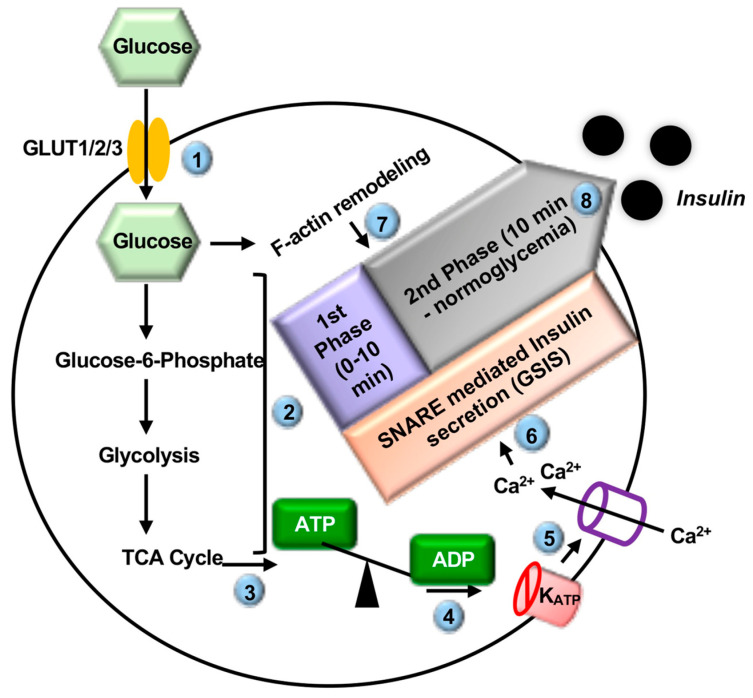
Type 2 diabetes (T2D) is one of the prominent causes of morbidity and mortality in the United States and beyond, reaching global pandemic proportions. One hallmark of T2D is dysfunctional glucose-stimulated insulin secretion from the pancreatic β-cell. Insulin is secreted via the recruitment of insulin secretory granules to the plasma membrane, where the soluble N-ethylmaleimide-sensitive factor attachment protein receptors (SNAREs) and SNARE regulators work together to dock the secretory granules and release insulin into the circulation. SNARE proteins and their regulators include the Syntaxins, SNAPs, Sec1/Munc18, VAMPs, and double C2-domain proteins. Recent studies using genomics, proteomics, and biochemical approaches have linked deficiencies of exocytosis proteins with the onset and progression of T2D. Promising results are also emerging wherein restoration or enhancement of certain exocytosis proteins to β-cells improves whole-body glucose homeostasis, enhances β-cell function, and surprisingly, protection of β-cell mass. Intriguingly, overexpression and knockout studies have revealed novel functions of certain exocytosis proteins, like Syntaxin 4, suggesting that exocytosis proteins can impact a variety of pathways, including inflammatory signaling and aging. In this review, we present the conventional and unconventional functions of β-cell exocytosis proteins in normal physiology and T2D and describe how these insights might improve clinical care for T2D.

Keywords: DOC2b; STX4; exocytosis; insulin secretion; β-cell function; β-cell mass; β-cell senescence/aging.


Conflict of interest statement

The authors declare no conflict of interest.

Figures

Figure 1
Figure 1
The steps of glucose stimulated insulin secretion (GSIS). Glucose enters the pancreatic β-cell via the GLUT1 or 3 (human)/GLUT2 (rodent) transporter (❶) and is rapidly metabolized via glycolysis and the tricarboxylic acid (TCA) cycle (❷). This increases the ATP/ADP ratio (❸), thereby closing the plasma membrane (PM)-localized ATP sensitive potassium channels (KATP) (❹), resulting in depolarization of the PM, opening of voltage-sensitive Ca2+ channels at the PM (❺), and influx of Ca2+ into the β-cell. Increased Ca2+ (❻), as well as glucose-induced F-actin remodeling (❼), results in soluble N-ethylmaleimide-sensitive factor attachment protein receptor (SNARE)-mediated fusion of insulin secretory granules to the PM and biphasic insulin release from the β-cell (❽). Steps are indicated by (circled numbers).
Figure 2
Figure 2
Role of STX1 in regulating insulin secretory granule (ISG) Pools and GSIS. During first–phase GSIS (0–10 min following glucose entry) (❶), three SNARE proteins: The STX1 open form (STX1OP), SNAP23/25, and VAMP2, assemble to form a heterotrimeric SNARE complex (❷) in the β-cell. This leads to fusion of the ISGs with the plasma membrane and release of insulin cargo into the extracellular space (❸). STX1 positively regulates the readily releasable pool (RRP) and newcomer pool of ISGs in the β-cell. Overexpression of STX1 decreases Ca2+ channel activity by physically interacting with it and thereby reduces GSIS. Blue arrows depict ISG movements, “+” arrows depict positive regulatory roles, “-“ arrow depicts a negative regulatory role. Steps are indicated by (circled numbers).
Figure 3
Figure 3
Roles of Syntaxin 4 in regulating ISG Pools and GSIS. (A) During the first (glucose stimulation 0–10min) (❶) and (B) second phases (glucose stimulation >10 min) (❺) of GSIS, (A, B) glucose stimulation tyrosine phosphorylates Munc18c and tyrosine phosphorylated Munc18c transiently switches its binding from STX4 to DOC2b (❷, ❻) in the β-cell. This results in the transition of STX4 from its closed (STX4CL) to open (STX4OP) conformation and the assembly of the three SNARE proteins to form a heterotrimeric complex: STX4, SNAP25 or SNAP23 and VAMP2 (❸, ❼) in the β-cell. This leads to SNARE-mediated ISG fusion with the PM and release of insulin cargo into the extracellular space (❹, ❽). STX4 positively regulates the RRP of ISGs. STX4 also positively regulates ISG refilling or newcomer pool of ISGs, possibly via its direct interaction with F-actin in the β-cell. Extra STX4 increases the amplitude of both phases of GSIS and enhances glucose homeostasis in vivo. Blue arrows depict ISG movements, “+” arrows depict positive regulatory roles. Steps are indicated by (circled numbers).
Figure 4
Figure 4
Role of SNAREs and SNARE-associated proteins in F-actin remodeling and GSIS. In the β-cell glucose stimulation dissociates gelsolin from the STX4. This results in the transition of STX4 from its closed (STX4CL) to open (STX4OP) conformation and F-actin remodeling via direct interaction with the F-actin network (❶). F-actin remodeling increases ISG movement towards PM and SNARE-mediated insulin secretion. Glucose-induced activation of PAK1 facilitates F-actin remodeling and recruitment of ISGs to the PM to support the second phase of insulin release, via activation of Rac1, Raf-1, MEK1/2, and ERK1/2 (❷). Glucose stimulation activates ezrin-radixin-moesin (ERM) proteins and activated ERM translocate to the PM and positively regulates ISG docking via interacting with the F-actin network (❸). Hypothetical positive regulatory role of DOC2b in F-actin remodeling in the β-cell (❹). Blue arrows depict ISG movements, “+” arrows depict positive regulatory roles. F-actin remodeling branches are depicted by (circled numbers).
Figure 5
Figure 5
Timeline of human islet transcriptome analysis details from the diabetic and non-diabetic donors. Transcriptome profiling via microarray, GWAS and laser capture (LCM) analyses between 2006 and 2014 elucidated the first associations between human T2D genes and changes attributed to β-cell function. After 2016, analyses transitioned largely to single-cell RNA sequencing (seq), providing β-cell specific changes. [numbers] refer to the list of references.
Figure 6
Figure 6
Role of STX4 in modulating inflammatory signals in the β-cell. STX4 binds to IĸBβ and prevents IL-1β and TNFα-induced proteasomal degradation of IĸBβ, thereby reducing p50-NF-ĸB nuclear translocation and suppressing gene expression of T2D-associated chemokine ligands CXCL9 and CXCL10. “T” arrow and red “X” denote inhibitory functions.
Figure 7
Figure 7
Inverse association between the peak amplitude of first phase GSIS and human islet donor age. (R2 = 0.84, p = 0.0014). Non-diabetic human donor islets were cultured overnight upon arrival and hand-picked to eliminate non-islet debris. Islets were evaluated by perifusion analyses. First-phase insulin release/acute insulin release (AIR) was quantified in 8 sets of donor islets across a 40-year span of ages.

Similar articles

Cited by

References

    1. Aslamy A., Thurmond D.C. Exocytosis proteins as novel targets for diabetes prevention and/or remediation? Am. J. Physiol. Regul. Integr. Comp. Physiol. 2017;312:R739–R752. doi: 10.1152/ajpregu.00002.2017. - DOI - PMC - PubMed
    1. ADA’s Primary Care Advisory Group (PCAG) American Diabetes Association. Standards of Medical Care in Diabetes—2020 Abridged for Primary Care Providers. Clin. Diabetes. 2020;38:10–38. doi: 10.2337/cd20-as01. - DOI - PMC - PubMed
    1. Butler A.E., Janson J., Bonner-Weir S., Ritzel R., Rizza R.A., Butler P.C. Beta-cell deficit and increased beta-cell apoptosis in humans with type 2 diabetes. Diabetes. 2003;52:102–110. doi: 10.2337/diabetes.52.1.102. - DOI - PubMed
    1. Del Guerra S., Lupi R., Marselli L., Masini M., Bugliani M., Sbrana S., Torri S., Pollera M., Boggi U., Mosca F., et al. Functional and molecular defects of pancreatic islets in human type 2 diabetes. Diabetes. 2005;54:727–735. doi: 10.2337/diabetes.54.3.727. - DOI - PubMed
    1. Hanley S.C., Austin E., Assouline-Thomas B., Kapeluto J., Blaichman J., Moosavi M., Petropavlovskaia M., Rosenberg L. {beta}-Cell mass dynamics and islet cell plasticity in human type 2 diabetes. Endocrinology. 2010;151:1462–1472. doi: 10.1210/en.2009-1277. - DOI - PubMed

LinkOut - more resources