. 2021 Feb 12;22(4):1835.
doi: 10.3390/ijms22041835.

A Toxic Synergy between Aluminium and Amyloid Beta in Yeast

Affiliations

A Toxic Synergy between Aluminium and Amyloid Beta in Yeast

Jamieson B Mcdonald et al. Int J Mol Sci. .

Abstract

Alzheimer's disease (AD), the most prevalent, age-related, neurodegenerative disease, is associated with the accumulation of amyloid beta (Aβ) and oxidative stress. However, the sporadic nature of late-onset AD has suggested that other factors, such as aluminium may be involved. Aluminium (Al3+) is the most ubiquitous neurotoxic metal on earth, extensively bioavailable to humans. Despite this, the link between Al3+ and AD has been debated for decades and remains controversial. Using Saccharomyces cerevisiae as a model organism expressing Aβ42, this study aimed to examine the mechanisms of Al3+ toxicity and its interactions with Aβ42. S. cerevisiae cells producing Aβ42 treated with varying concentrations of Al3+ were examined for cell viability, growth inhibition, and production of reactive oxygen species (ROS). Al3+ caused a significant reduction in cell viability: cell death in yeast producing green fluorescent protein tagged with Aβ42 (GFP-Aβ42) was significantly higher than in cells producing green fluorescent protein (GFP) alone. Additionally, Al3+ greatly inhibited the fermentative growth of yeast producing GFP-Aβ42, which was enhanced by ferric iron (Fe3+), while there was negligible growth inhibition of GFP cells. Al3+- induced ROS levels in yeast expressing native Aβ42 were significantly higher than in empty vector controls. These findings demonstrate Al3+ has a direct, detrimental toxic synergy with Aβ42 that can be influenced by Fe3+, causing increased oxidative stress. Thus, Al3+ should be considered as an important factor, alongside the known characteristic hallmarks of AD, in the development and aetiology of the disease.

Keywords: Alzheimer’s disease; Fenton chemistry; aluminium; amyloid beta; iron; oxidative stress; yeast.


Conflict of interest statement

The authors declare no conflict of interest.

Figures

Figure 1
Figure 1
The biological relationships and impacts Al3+ has on the characteristic hallmarks of Alzheimer’s disease (AD) (amyloid beta, biometal dyshomeostasis, and oxidative stress). Al3+ has been experimentally shown to induce all key pathological events associated with AD, at multiple levels.
Figure 2
Figure 2
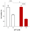
Al3+-mediated cell killing of Saccharomyces cerevisiae transformant strains BY4743 [p416GPD.GFP] (white bar) and BY4743 [p416GPD.GFPAβ] (red bar). S. cerevisiae transformant strains BY4743 [p416GPD.GFP] and BY4743 [p416GPD.GFPAβ] were suspended in water and treated with 10 mM Al2(SO4)3. After 24 h, cells were plated on yeast extract peptone dextrose (YEPD) and incubated for 4 days at 30 °C to determine cell viability. Values are from triplicates; the mean and standard deviation are shown. Values significantly different from 0 mM Al3+ and between the two transformant strains in a two-way ANOVA with Tukey’s post hoc analysis are indicated with asterisks: * p < 0.01, *** p < 0.0003, **** p < 0.0001.
Figure 3
Figure 3
Growth of S. cerevisiae BY4743 [p416GPD.GFP] and BY4743 [p416GPD.GFPAβ] transformants on low-pH and low-phosphate (LPP) medium containing varying concentrations of Al3+ incubated at 30 °C for 7 days. Analysis of growth inhibition was performed in triplicate rows (transformant strains) and compared. The difference in growth inhibition between the two transformants provides another line of evidence that the combination of Aβ42 and Al3+ has a synergistic toxicity towards cells, with Al3+ having a dose-dependent toxicity.
Figure 4
Figure 4
Growth of S. cerevisiae BY4743 [p416GPD.GFP] and BY4743 [p416GPD.GFP.Aβ] cells on LPP medium containing 1.6 mM of Al3+ and 2 mM Fe3+ indicated by (+), plates were incubated at 30 °C for 7 days.
Figure 5
Figure 5
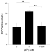
Al3+ induced reactive oxygen species (ROS) generation in S. cerevisiae BY4743 [pYEX.Aβ] (blue bar) and BY4743 [pYEX.BX] (green bar) using 2,7-dichlorodihydrofluorescein diacetate (H2DCFDA) staining. Values of dichlorofluorescein (DCF)-positive cell counts after 5 mM Al3+ treatment are from triplicates; the mean and standard deviation are shown. Values significantly different from 0 mM Al3+ and between the two transformant strains in a two-way ANOVA with Tukey’s post hoc analysis are indicated with asterisks: * p < 0.0312, ** p < 0.0074, *** p < 0.0005.
Figure 6
Figure 6
Glutathione (GSH) rescue of Al3+-induced ROS generation in S. cerevisiae BY4743 [pYEX.Aβ] using 2,7-dichlorodihydrofluorescein diacetate (H2DCFDA) staining. Bars represent dichlorofluorescein (DCF)-positive cell counts after 5 mM Al3+ treatment, 5 mM GSH was used to rescue cells from oxidative stress caused by treatment with 5 mM Al3+ and the presence of Aβ42. Values are from triplicates; the mean and standard deviation are shown. Values significantly different from 0 mM, 5 mM Al3+ and GSH rescue in a one-way ANOVA with Tukey’s post hoc analysis are indicated with asterisks: ** p < 0.0040.

References

    1. Tomljenovic L. Aluminum and Alzheimer’s Disease: After a century of controversy, is there a plausible link? J. Alzheimer’s Dis. 2011;23:567–598. doi: 10.3233/JAD-2010-101494. - DOI - PubMed
    1. Kawahara M., Kato-Negishi M. Link between aluminum and the pathogenesis of Alzheimer’s Disease: The integration of the aluminum and amyloid cascade hypotheses. Int. J. Alzheimer’s. 2011;2011:1–17. doi: 10.4061/2011/276393. - DOI - PMC - PubMed
    1. Rondeau V., Commenges D., Jacqmin-Gadda H., Dartigues, J-F Relation between aluminum concentrations in drinking water and Alzheimer’s Disease: An 8-year follow-up study. Am. J. Epidemiol. 2000;152:59–66. doi: 10.1093/aje/152.1.59. - DOI - PMC - PubMed
    1. Li Y., Jiao Q., Xu H., Du X., Shi L., Jia F., Jiang H. Biometal dyshomeostasis and toxic metal accumulations in the development of Alzheimer’s Disease. Front. Mol. Neurosci. 2017;10:339. doi: 10.3389/fnmol.2017.00339. - DOI - PMC - PubMed
    1. Yokel R.A. Blood-brain barrier flux of aluminum, manganese, iron and other metals suspected to contribute to metal-induced neurodegeneration. J. Alzheimer’s Dis. 2006;10:223–253. doi: 10.3233/JAD-2006-102-309. - DOI - PubMed

MeSH terms

LinkOut - more resources