Review
. 2021 Feb 12;22(4):1849.
doi: 10.3390/ijms22041849.

Insulin-Like Growth Factor 2 As a Possible Neuroprotective Agent and Memory Enhancer-Its Comparative Expression, Processing and Signaling in Mammalian CNS

Affiliations
Review

Insulin-Like Growth Factor 2 As a Possible Neuroprotective Agent and Memory Enhancer-Its Comparative Expression, Processing and Signaling in Mammalian CNS

Alexander Beletskiy et al. Int J Mol Sci. .

Abstract

A number of studies performed on rodents suggest that insulin-like growth factor 2 (IGF-2) or its analogs may possibly be used for treating some conditions like Alzheimer's disease, Huntington's disease, autistic spectrum disorders or aging-related cognitive impairment. Still, for translational research a comparative knowledge about the function of IGF-2 and related molecules in model organisms (rats and mice) and humans is necessary. There is a number of important differences in IGF-2 signaling between species. In the present review we emphasize species-specific patterns of IGF-2 expression in rodents, humans and some other mammals, using, among other sources, publicly available transcriptomic data. We provide a detailed description of Igf2 mRNA expression regulation and pre-pro-IGF-2 protein processing in different species. We also summarize the function of IGF-binding proteins. We describe three different receptors able to bind IGF-2 and discuss the role of IGF-2 signaling in learning and memory, as well as in neuroprotection. We hope that comprehensive understanding of similarities and differences in IGF-2 signaling between model organisms and humans will be useful for development of more effective medicines targeting IGF-2 receptors.

Keywords: CNS; IGF-binding proteins; IGF1R; IGF2R/CI-M6P; insulin receptor; insulin-like growth factor 2; learning and memory; neuroprotection; species-specific gene expression.


Conflict of interest statement

The authors declare no conflict of interest.

Figures

Figure 1
Figure 1
Insulin-like growth factor 2 (IGF-2) expression levels in different organs during postnatal development in rats according to Gene Expression Omnibus data repository [57,58]. Age is given in days. LogFPKM—logarithm of FPKM (fragments per kilobase of exon model per million reads mapped) value.
Figure 2
Figure 2
Levels of Igf2 mRNA in the choroid plexus of rats of different age according to Gene Expression Omnibus data repository. (a) Igf2 expression level in the choroid plexus from embryonal day 19 (E19), postnatal day 2 (P2) and adult rat according to the data from [63,64]. FPKM (fragments per kilobase of exon model per million reads mapped); (b) Igf2 expression level in the choroid plexus from embryonal day 15 (E15) and adult rat according to the data from [65,66].
Figure 3
Figure 3
Cell clusters of the mouse brain showing the highest expression of Igf2 mRNA after single-cell sequencing from 9 mouse brain regions. The figure was generated by DropViz [68,69] and slightly modified. 1 It was noted that small fractions of choroid plexus were sampled from ventricle-adjacent regions including hippocampus [69].
Figure 4
Figure 4
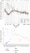
IGF2 mRNA expression levels in human cortical regions during pre- and postnatal development. (a) IGF2 mRNA expression levels in neocortical areas assessed in the Human Brain Transcriptome project [76,77]. OFC—orbital prefrontal cortex, DFC—dorsolateral prefrontal cortex, VFC—ventrolateral prefrontal cortex, MFC—medial prefrontal cortex, M1C—primary motor (M1) cortex, S1C—primary somatosensory (S1) cortex, IPC—posterior inferior parietal cortex, A1C—primary auditory (A1) cortex, STC—superior temporal cortex, ITC—inferior temporal cortex, V1C—primary visual (V1) cortex. Vertical line denotes the time of birth. Age is given in postconceptional days. (b) IGF2 mRNA expression levels (RPKM, reads per kilobase of exon model per million mapped reads) in dorsolateral prefrontal cortex during human development. Image is based on the data obtained from [78,79]. pcw—postconceptional week, mos—months of postnatal development, yrs—donor age in years.
Figure 5
Figure 5
IGF2 mRNA expression in human choroid plexus according to [85,86]. Microarray data (median-centered values, normalized and cross-comparable across replicates) were processed using the GEO2R tool [87,88]. (a) IGF2 expression levels in choroid plexus samples from individual donors. Con—control (healthy) donors, AD_V-VI—advanced Alzheimer’s disease (Braak stage V–VI), AD_III-IV—Alzheimer’s disease (Braak III–IV), HD—Huntington’s disease, FTD—frontotemporal dementia, FTD_MN—frontotemporal dementia and motor neuron disease, FTD_Pick—Pick’s disease (a specific type of FTD); (b) Averaged density distribution of gene expression in choroid plexus from healthy donors. Red dash lines indicate minimal and maximal IGF2 expression levels. IGF2 gene is located in the far right part of the graph, meaning that IGF2 is one of the most expressed among all the studied genes. (Analysis of density distribution of gene expression is described in detail in [89]).
Figure 6
Figure 6
Currently known IGF-2 proteolysis sites and IGF-2 proteoforms discovered in blood serum and brain samples of different species. (a) According to various reports, pro-IGF-2 (1-156 aa) has a molecular weight about 20–26 kDa in different tissues [36,107,108]. (b) Proteoforms 1–87 aa and 1–104 aa, also known as “big IGF-2”. A few products of incomplete pro-IGF-2 cleavage weighing from 8 to 18 kDa were registered in some experiments [35,36,106,112]. (c) Mature IGF-2 (1-67 aa) has a molecular weight of 7.5 kDa [7,54,106,109,110]. (d) 15 and 20 kDa IGF-2 proteoforms were found in rat dorsal hippocampus [108]. (e) 14 and 21–22 kDa IGF-2 proteoforms were detected in mouse brain regions (hippocampus, cortex, striatum and cerebellum), with some additional bands being present on Western blots [111]. 17 kDa IGF-2 was found in mouse hippocampus and entorhinal cortex [20]. (f) Mature IGF-2, “big IGF-2” and pro-IGF-2 are present in the serum of neonatal [54] and 10 days old rats [112]. (g) In the serum of adult rats, pro-IGF-2 is the predominant proteoform, while mature and “big” IGF-2 are either present in low quantities [54] or not detected at all [112]. (h) Mature IGF-2 (7.5 kDa) is the main IGF-2 proteoform in adult human serum (plasma) [54,110,112], while “big IGF-2” is present there in low quantity [110,112]. Pro-IGF-2 was also revealed in the human serum in some studies [112] but not detected in others [54,110]. Thick lines denote proteoforms that were clearly present in samples, thin lines denote proteoforms that were reported to be present in low quantity, dash lines denote proteoforms that were detected only in some of all the referenced studies.
Figure 7
Figure 7
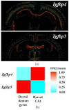
Expression patterns of Igfbp4 and Igfbp5 mRNA in the mouse brain according to databases. (a) In situ hybridization results from Allen Mouse Brain Atlas [144,145,146]; (b) Normalized Igfbp4 and Igfbp5 expression heatmap for dorsal CA1 and dentate gyrus hippocampal principal neurons from Hippocampus RNA-seq atlas [147,148].

Similar articles

Cited by

References

    1. Bracko O., Singer T., Aigner S., Knobloch M., Winner B., Ray J., Clemenson G.D., Jr., Suh H., Couillard-Despres S., Aigner L., et al. Gene expression profiling of neural stem cells and their neuronal progeny reveals IGF2 as a regulator of adult hippocampal neurogenesis. J. Neurosci. 2012;32:3376–3387. doi: 10.1523/JNEUROSCI.4248-11.2012. - DOI - PMC - PubMed
    1. Martín-Montañez E., Millon C., Boraldi F., Garcia-Guirado F., Pedraza C., Lara E., Santin L.J., Pavia J., Garcia-Fernandez M. IGF-II promotes neuroprotection and neuroplasticity recovery in a long-lasting model of oxidative damage induced by glucocorticoids. Redox Biol. 2017;13:69–81. doi: 10.1016/j.redox.2017.05.012. - DOI - PMC - PubMed
    1. Russo V.C., Gluckman P.D., Feldman E.L., Werther G.A. The Insulin-Like Growth Factor System and Its Pleiotropic Functions in Brain. Endocr. Rev. 2005;26:916–943. doi: 10.1210/er.2004-0024. - DOI - PubMed
    1. Belfiore A., Malaguarnera R., Vella V., Lawrence M.C., Sciacca L., Frasca F., Morrione A., Vigneri R. Insulin Receptor Isoforms in Physiology and Disease: An Updated View. Endocr. Rev. 2017;38:379–431. doi: 10.1210/er.2017-00073. - DOI - PMC - PubMed
    1. Yakar S., Liu J.L., Stannard B., Butler A., Accili D., Sauer B., LeRoith D. Normal growth and development in the absence of hepatic insulin-like growth factor I. Proc. Natl. Acad. Sci. USA. 1999;96:7324–7329. doi: 10.1073/pnas.96.13.7324. - DOI - PMC - PubMed