Review
. 2021 Feb 27;13(5):986.
doi: 10.3390/cancers13050986.

Oxidative Stress in the Tumor Microenvironment and Its Relevance to Cancer Immunotherapy

Affiliations
Review

Oxidative Stress in the Tumor Microenvironment and Its Relevance to Cancer Immunotherapy

Nada S Aboelella et al. Cancers (Basel). .

Abstract

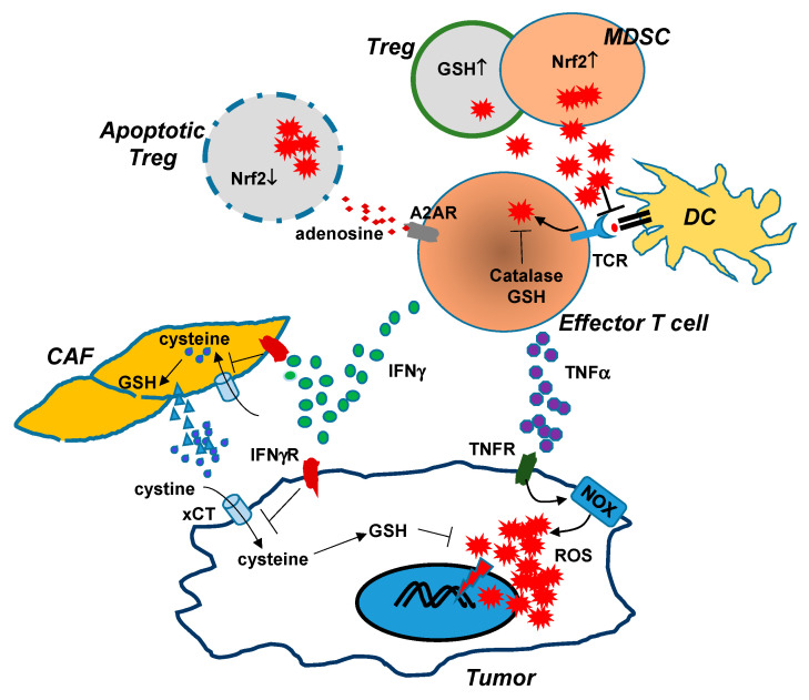
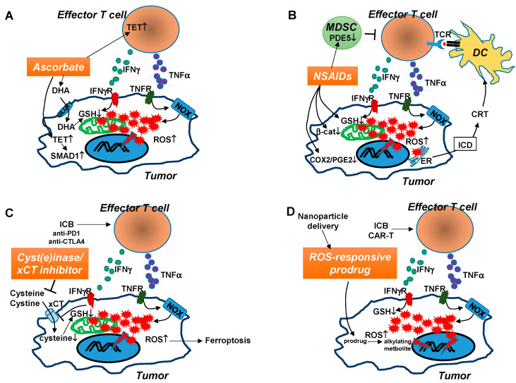
It has been well-established that cancer cells are under constant oxidative stress, as reflected by elevated basal level of reactive oxygen species (ROS), due to increased metabolism driven by aberrant cell growth. Cancer cells can adapt to maintain redox homeostasis through a variety of mechanisms. The prevalent perception about ROS is that they are one of the key drivers promoting tumor initiation, progression, metastasis, and drug resistance. Based on this notion, numerous antioxidants that aim to mitigate tumor oxidative stress have been tested for cancer prevention or treatment, although the effectiveness of this strategy has yet to be established. In recent years, it has been increasingly appreciated that ROS have a complex, multifaceted role in the tumor microenvironment (TME), and that tumor redox can be targeted to amplify oxidative stress inside the tumor to cause tumor destruction. Accumulating evidence indicates that cancer immunotherapies can alter tumor redox to intensify tumor oxidative stress, resulting in ROS-dependent tumor rejection. Herein we review the recent progresses regarding the impact of ROS on cancer cells and various immune cells in the TME, and discuss the emerging ROS-modulating strategies that can be used in combination with cancer immunotherapies to achieve enhanced antitumor effects.

Keywords: immunotherapy; oxidative stress; reactive oxygen species; tumor microenvironment.


Conflict of interest statement

The authors declare no conflict of interest.

Figures

Figure 1
Figure 1
Interactions between cells in the TME lead to changes in redox status. Tumor-reactive effector T cells (CD4+ and CD8+) can induce increased levels of ROS in cancer cells via the actions of IFNγ and TNFα. TNFα signaling in tumor cells activates NADPH oxidases, which lead to increased production of ROS. IFNγ signaling in tumor cells diminishes xCT expression through transcriptional inhibition, reducing tumor uptake of extracellular cystine and subsequent GSH synthesis. IFNγ signaling in cancer-associated fibroblasts (CAF) blocks the release of GSH and cysteine, further depleting the extracellular pool of cystine and cysteine available to cancer cells. The severe redox imbalance in cancer cells, caused by TNFα-driven ROS production and IFNγ-induced GSH deficiency, leads to extensive oxidative damages and eventual tumor cell death. However, effector T cells are also susceptible to oxidative stress in the TME. ROS induced upon TCR engagement are counterbalanced by increased antioxidant systems such as GSH and catalase. T cell dysfunction may occur when effector T cells are exposed to ROS produced by MDSCs and Tregs, which are more resistant to oxidative stress due to their increased antioxidant systems. High levels of extracellular ROS can disrupt antigen-presentation between T cells and DCs, and can affect tumor antigen recognition by T cells. Some apoptosis-prone Tregs can increase the presence of adenosine in the TME, which suppresses the function of effector T cells in an A2AR-dependent manner. Therapeutic interventions should be directed to enhance T cell-induced tumor oxidative stress while enabling T cells to resist the elevated oxidative stress in the TME.
Figure 2
Figure 2
Hypothetical mechanisms by which certain pro-oxidants enhance the efficacy of cancer immunotherapies. Tumor-reactive effector T cells, emerging in the TME after either ICB therapy or adoptive transfer, can mediate tumor killing via well-characterized mechanisms involving cytolytic granules such as perforin and granzymes, and apoptosis-inducing ligands such as FASL and TRAIL. Increasing evidence reveals that inflammatory cytokines produced by effector T cells, including TNFα and IFNγ, can exert antitumor effect by modulating tumor redox. TNFα signaling in cancer cells activates NOX-dependent ROS production, while IFNγ signaling exacerbates GSH deficiency by suppressing the cysteine/glutamate transporter xCT. The combined effects of TNFα and IFNγ lead to substantial ROS accumulation in tumor cells, rendering them vulnerable to further redox disruption which can be incited by a pro-oxidant. A suitable pro-oxidant should preferentially induce oxidative stress in cancer cells without harming antitumor T cells. The potential mechanisms of action of four immunotherapy-compatible pro-oxidants are illustrated. (A). Pharmacological dose of ascorbate, in its oxidized form DHA, can be preferentially taken into cancer cells via the glucose transporter (GLUT1). Intracellular DHA is reduced to ascorbate at the expense of GSH. The GSH shortage aggravates ROS accumulation, which damages DNA/protein/lipid and derails cell metabolism, leading to tumor cell death. Meanwhile, ascorbate may induce TET activities in antitumor T cells and tumor cells. TET activation in T cells leads to enhanced function of T cells through epigenetic modifications. TET activation in tumor cells results in demethylation and activation of SMAD1, which increases tumor chemosensitivity. (B). NSAIDs can act as pro-oxidants to reduce GSH and thereby increase the levels of ROS in tumor cells. NSAID-induced ER stress may lead to release of calreticulin (CRT), a DAMP molecule characteristic of ICD, which can attract and activate DCs, which in turn elicit antitumor CD8+ T cell responses. In addition, NSAIDs can suppress tumor cell growth by its inhibitory effect on β-catenin, COX2, and PGE2. Some NSAIDs may reduce MDSC activity by inhibiting PDE5 function in MDSCs. (C). Cyst(e)inase or xCT inhibitors can reduce the presence or block the uptake of extracellular cystine and cysteine, which tumor cells rely on to synthesize GSH, respectively. Cyst(e)inase can act in concert with ICB-induced antitumor T cells to drive tumor cell ferroptosis. (D). ROS-responsive prodrugs can be effectively delivered to tumor loci by nanoparticles. The increased levels of ROS in the TME can activate these prodrugs, which give rise to alkylating metabolites to cause further DNA damage and intensify oxidative stress in tumor cells. These prodrugs may synergize with antitumor T cells because ROS accumulated in tumor cells after immunotherapies such as ICB or CAR-T therapy can effectively activate prodrugs, which in turn further amplify ROS in tumor cells to drive apoptosis.

References

    1. Schumacker P.T. Reactive oxygen species in cancer cells: Live by the sword, die by the sword. Cancer Cell. 2006;10:175–176. doi: 10.1016/j.ccr.2006.08.015. - DOI - PubMed
    1. Liou G.Y., Storz P. Reactive oxygen species in cancer. Free Radic. Res. 2010;44:479–496. doi: 10.3109/10715761003667554. - DOI - PMC - PubMed
    1. Chio I.I.C., Tuveson D.A. ROS in Cancer: The Burning Question. Trends Mol. Med. 2017;23:411–429. doi: 10.1016/j.molmed.2017.03.004. - DOI - PMC - PubMed
    1. Reczek C.R., Chandel N.S. The Two Faces of Reactive Oxygen Species in Cancer. Annu. Rev. Cancer Biol. 2017;1:79–98. doi: 10.1146/annurev-cancerbio-041916-065808. - DOI
    1. Murphy M.P. How mitochondria produce reactive oxygen species. Biochem. J. 2009;417:1–13. doi: 10.1042/BJ20081386. - DOI - PMC - PubMed