Review
. 2021 Feb 27;22(5):2362.
doi: 10.3390/ijms22052362.

MicroRNAs Modulate Signaling Pathways in Osteogenic Differentiation of Mesenchymal Stem Cells

Affiliations
Review

MicroRNAs Modulate Signaling Pathways in Osteogenic Differentiation of Mesenchymal Stem Cells

Chiara Mazziotta et al. Int J Mol Sci. .

Abstract

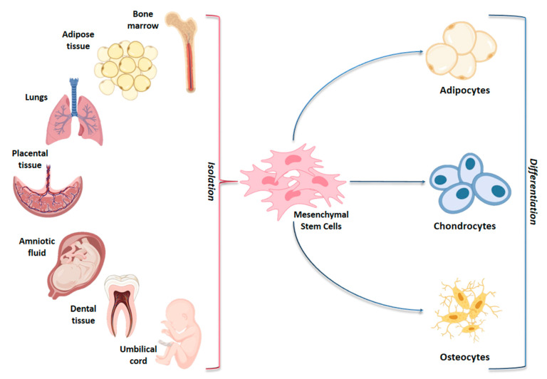
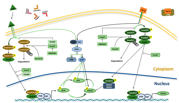
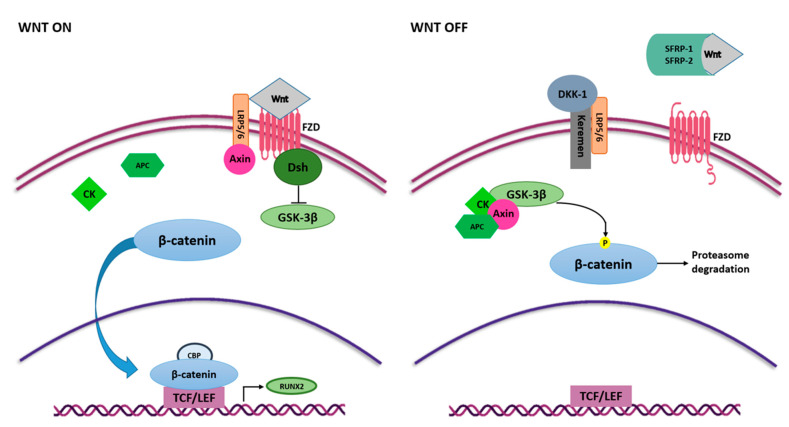
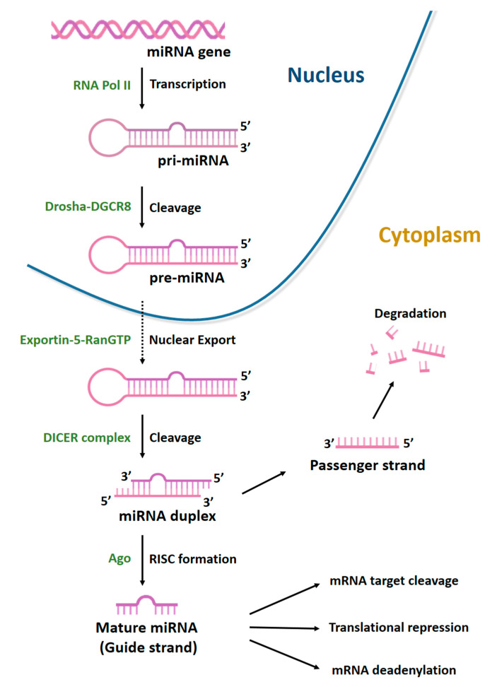
Mesenchymal stem cells (MSCs) have been identified in many adult tissues and they have been closely studied in recent years, especially in view of their potential use for treating diseases and damaged tissues and organs. MSCs are capable of self-replication and differentiation into osteoblasts and are considered an important source of cells in tissue engineering for bone regeneration. Several epigenetic factors are believed to play a role in the osteogenic differentiation of MSCs, including microRNAs (miRNAs). MiRNAs are small, single-stranded, non-coding RNAs of approximately 22 nucleotides that are able to regulate cell proliferation, differentiation and apoptosis by binding the 3' untranslated region (3'-UTR) of target mRNAs, which can be subsequently degraded or translationally silenced. MiRNAs control gene expression in osteogenic differentiation by regulating two crucial signaling cascades in osteogenesis: the transforming growth factor-beta (TGF-β)/bone morphogenic protein (BMP) and the Wingless/Int-1(Wnt)/β-catenin signaling pathways. This review provides an overview of the miRNAs involved in osteogenic differentiation and how these miRNAs could regulate the expression of target genes.

Keywords: MSCs; bone; bone regeneration; cell differentiation; mesenchymal stem cells; miRNA; miRNAome; microRNA; osteogenesis; osteogenic differentiation; stem cells.


Conflict of interest statement

The authors declare no conflict of interest.

Figures

Figure 1
Figure 1
Schematic representation of adult and fetal/neonatal tissue sources of mesenchymal stem cells (MSCs) and their potential of differentiation in various cell lines. MSCs can be isolated from several tissue sources in the body and they may differentiate into adipocytes, chondrocytes and osteocytes.
Figure 2
Figure 2
BMPs and TGF-β signaling pathways in osteogenesis. Interaction between TGF-βs and BMPs and receptors initiate SMAD-dependent and non-SMAD-dependent signaling pathways. In SMAD-dependent signaling pathway, interaction between TGF-β and receptor leads to SMAD2/3 (R-SMAD) phosphorylation. Phosphorylated R-SMAD (SMAD2/3-Pi) binds SMAD4 protein and migrates to the nucleus of the cell where, alongside P300 and CREB-binding protein (CBP) coactivators, it controls RUNX2 expression. In the nucleus, SMAD2/3-Pi without SMAD4 recruits HDAC4 and HDAC5 and blocks RUNX2 activity. In BMP signaling, R-SMAD (SMAD1/5/8) binds SMAD4 and migrates to the nucleus, inducing RUNX2 and OSX expression through DLX5 activation. In the non-SMAD-dependent signaling pathway, TAK1 and TAB1 activate the MKKs p38 MAPK or Erk cascades, inducing DLX5, RUNX2 and OSX phosphorylation. The interaction between TGF-β and its receptor is blocked by the latent TGF-β binding protein (LTBP), which binds to TGF-β. Similarly, in order to prevent the BMP–receptor interaction, Gremlin, Chordin, Follistatin and Noggin proteins bind to BMP. The translocation of SMAD2/3 and SMAD1/5/8 into the nucleus is prevented by SMAD7 that, along with SMURF1/2, induce their proteasome-mediated degradation. In addition, the binding between ARKADIA and SMAD6/7 proteins positively regulates osteoblast differentiation.
Figure 3
Figure 3
Wnt/β-catenin signaling pathway in osteogenesis. Wnt ligands interact with FZD and activate Wnt signaling pathway. FZD binds Dsh that inhibits GSK3-β activity, and thus the phosphorylation of β-catenin. β-catenin translocates into the nucleus, where it binds to TCF/LEF and CBP and induces the expression of RUNX2. Interaction between Wnt ligands and SFRP-1/2 and the binding of LRP5/6 and Keremen with DKK-1 antagonist switch off Wnt signaling pathway. In this case, GSK3-β, together with Axin, CK and APC, phosphorylate β-catenin, inducing proteasome-mediated degradation.
Figure 4
Figure 4
Biogenesis of microRNAs. MiRNAs are transcribed by RNA polymerase II (Pol II) to generate a long precursor transcript named primary microRNA (pri-miRNA). This RNA molecule folds up into a secondary structure (stem-loop) to form a partial double helix, composed of 100–1000 nt with a 5′-cap. MiRNA maturation process can be divided into three phases. In the first, known as cropping, pri-miRNA is converted into the precursor miRNA (pre-miRNA) via the cutting activity of the Drosha enzyme, a nuclear endoribonuclease III. Pre-miRNA has a hairpin structure (stem-loop) and a length of about 70–80 nt. Following cropping, the pre-miRNA has a 5′P and a 3′OH and 2–3 nt at the protruding end with a single strand. In the second phase, the nuclear export factors Exportin-5 and ras-related nuclear protein (RAN-GTP) mediate the export of pre-miRNAs from nucleus to cytoplasm. In the third phase, known as dicing, another type of RNA endonuclease III, known as Dicer, processes pre-miRNA in the cytoplasm by cleaving it into an 18–22 nt double-stranded miRNA (miRNA duplex). Lastly, mature miRNA incorporated into the RNA-induced silencing complex (RISC) is able to bind the 3′UTR region of its target mRNA.

Similar articles

Cited by

References

    1. Chen Q., Shou P., Zheng C., Jiang M., Cao G., Yang Q., Cao J., Xie N., Velletri T., Zhang X., et al. Fate Decision of Mesenchymal Stem Cells: Adipocytes or Osteoblasts? Cell Death Differ. 2016;23:1128–1139. doi: 10.1038/cdd.2015.168. - DOI - PMC - PubMed
    1. Mazzoni E., D’Agostino A., Iaquinta M.R., Bononi I., Trevisiol L., Rotondo J.C., Patergnani S., Giorgi C., Gunson M.J., Arnett G.W., et al. Hydroxylapatite-Collagen Hybrid Scaffold Induces Human Adipose-Derived Mesenchymal Stem Cells to Osteogenic Differentiation in Vitro and Bone Regrowth in Patients. Stem Cells Transl. Med. 2020;9:377–388. doi: 10.1002/sctm.19-0170. - DOI - PMC - PubMed
    1. Friedenstein A.J., Petrakova K.V., Kurolesova A.I., Frolova G.P. Heterotopic of Bone Marrow. Analysis of Precursor Cells for Osteogenic and Hematopoietic Tissues. Transplantation. 1968;6:230–247. doi: 10.1097/00007890-196803000-00009. - DOI - PubMed
    1. Mortada I., Mortada R. Epigenetic Changes in Mesenchymal Stem Cells Differentiation. Eur. J. Med. Genet. 2018;61:114–118. doi: 10.1016/j.ejmg.2017.10.015. - DOI - PubMed
    1. Iaquinta M.R., Mazzoni E., Bononi I., Rotondo J.C., Mazziotta C., Montesi M., Sprio S., Tampieri A., Tognon M., Martini F. Adult Stem Cells for Bone Regeneration and Repair. Front. Cell Dev. Biol. 2019;7:268. doi: 10.3389/fcell.2019.00268. - DOI - PMC - PubMed

LinkOut - more resources