. 2021 Feb 27;13(3):374.
doi: 10.3390/v13030374.

Analysis of a Novel Bacteriophage vB_AchrS_AchV4 Highlights the Diversity of Achromobacter Viruses

Affiliations

Analysis of a Novel Bacteriophage vB_AchrS_AchV4 Highlights the Diversity of Achromobacter Viruses

Laura Kaliniene et al. Viruses. .

Abstract

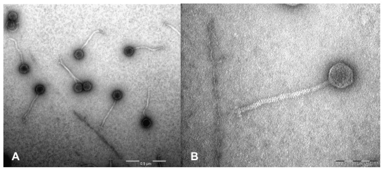
Achromobacter spp. are ubiquitous in nature and are increasingly being recognized as emerging nosocomial pathogens. Nevertheless, to date, only 30 complete genome sequences of Achromobacter phages are available in GenBank, and nearly all of those phages were isolated on Achromobacter xylosoxidans. Here, we report the isolation and characterization of bacteriophage vB_AchrS_AchV4. To the best of our knowledge, vB_AchrS_AchV4 is the first virus isolated from Achromobacter spanius. Both vB_AchrS_AchV4 and its host, Achromobacter spanius RL_4, were isolated in Lithuania. VB_AchrS_AchV4 is a siphovirus, since it has an isometric head (64 ± 3.2 nm in diameter) and a non-contractile flexible tail (232 ± 5.4). The genome of vB_AchrS_AchV4 is a linear dsDNA molecule of 59,489 bp with a G+C content of 62.8%. It contains no tRNA genes, yet it includes 82 protein-coding genes, of which 27 have no homologues in phages. Using bioinformatics approaches, 36 vB_AchrS_AchV4 genes were given a putative function. A further four were annotated based on the results of LC-MS/MS. Comparative analyses revealed that vB_AchrS_AchV4 is a singleton siphovirus with no close relatives among known tailed phages. In summary, this work not only describes a novel and unique phage, but also advances our knowledge of genetic diversity and evolution of Achromobacter bacteriophages.

Keywords: Achromobacter; Siphoviridae; bacteriophage.


Conflict of interest statement

The authors declare no conflict of interest. The funders had no role in the design of the study; in the collection, analyses, or interpretation of data; in the writing of the manuscript, or in the decision to publish the results.

Figures

Figure 1
Figure 1
Electron micrographs of negatively stained AchV4 particles. (A) TEM micrograph showing CsCl-purified AchV4 virions (the scale bar corresponds to 0.5 μm); (B) a close-up view of an AchV4 particle (the scale bar corresponds to 0.1 μm).
Figure 2
Figure 2
Functional genome map of bacteriophage AchV4. The coding capacity of the AchV4 genome is shown. Functions were assigned based on homology to known genes and/or MS-MS analysis. The color code is as follows: blue, structural proteins and those involved in virion morphogenesis; light blue, terminase complex; yellow, DNA metabolism; orange, transcription; violet/purple, lysis; green, lysogeny; red, genes that encode unique proteins with no reliable homology to database entries; grey, genes of unknown function. Pm—promoter. The figure was generated using Geneious Prime 2021.01.
Figure 3
Figure 3
Phylogenetic analysis. Neighbor-joining tree analysis based on the ClustalW alignment of: the large terminase subunit (A) and the major capsid protein (B) sequences. The percentage of replicate trees in which the associated taxa clustered together in the bootstrap test (500 replicates) is shown next to the branches. Evolutionary analyses were conducted in MEGA X. US—unclassified siphovirus; UM—unclassified myovirus; UP—unclassified podovirus; S, Sa—Siphoviridae, Sanovirus; S, Chi—Siphoviridae, Chivirus; S, Ahd—Siphoviridae, Ahduovirus; S, Nazg—Siphoviridae; Nazgulvirus; M, Ou—Myoviridae; Ounavirinae; AchV4 is indicated by a red dot.
Figure 4
Figure 4
Phylogenetic analysis. Neighbor-joining tree analysis based on the ClustalW alignment of: DNA polymerase (A) and phage endolysin (B) sequences. The percentage of replicate trees in which the associated taxa clustered together in the bootstrap test (500 replicates) is shown next to the branches. Evolutionary analyses were conducted in MEGA X. US—unclassified siphovirus; UM—unclassified myovirus; UP—unclassified podovirus; S, Seu—Siphoviridae, Seuratvirus; S, No—Siphoviridae, Nonagvirus; S, Nip—Siphoviridae, Nipunavirus; S, Kil—Siphoviridae, Kilunavirus; S, Sep—Siphoviridae, Septimatrevirus M, Ota—Myoviridae; Otagovirus; M, Pak—Myoviridae, Pakpunavirus; M, Phi—Myoviridae, Phikzvirus; M, Kis—Myoviridae, Kisquattuordecimvirus; M, Are—Myoviridae, Aresaunavirus; M, Ci—Myoviridae, Citexvirus; M, No—Myoviridae, Noxifervirus; A, Li—Ackermannviridae, Limestonevirus; A, Ag—Ackermannviridae, Agtrevirus; A, Ku—Ackermannviridae, Kuttervirus; A, Ta—Ackermannviridae, Taipeivirus; ND—not determined. AchV4 is indicated by a red dot.
Figure 5
Figure 5
Phylogenetic analysis based on genome-wide sequence similarities computed by tBLASTx. The proteomic tree based on the complete genome sequences of phages with SG values to AchV4 ranging from 0.1581 (Phobos) to 0.05 (Mycobacterium phage JAMaL). The figure was generated by ViPTree.
Figure 6
Figure 6
Genome alignment of AchV4, phiAxp-2, Phobos, PspYZU01, and YMC16/01/N133_KPN_BP. All tBLASTx alignments are represented by colored lines between two genomes. Color scale represents the tBLASTx percent identity. The figure was generated by ViPTree.
Figure 7
Figure 7
Splitstree representation of the unrooted proteomic tree generated by ViPTree web-service based on the genome-wide similarity relationships between Achromobacter bacteriophages. Purple, phages of the family Schitoviridae; red, siphoviruses that belong to the genus Steinhofvirus; blue, phages from the family Autographiviridae. UM—unclassified myovirus; MM—family Myoviridae, genus Mieseafarmvirus; US—unclassified siphoviruses.

Similar articles

Cited by

References

    1. Busse H.-J., Auling G. Achromobacter. In: Trujillo M.E., Dedysh S., DeVos P., Hedlund B., Kämpfer P., Rainey F.A., Whitman W.B., editors. Bergey’s Manual of Systematics of Archaea and Bacteria. John Wiley & Sons; Hoboken, NJ, USA: 2015. - DOI
    1. Swenson C.E., Sadikot R.T. Achromobacter respiratory infections. Ann. Am. Thorac. Soc. 2015;12:252–258. doi: 10.1513/AnnalsATS.201406-288FR. - DOI - PubMed
    1. Kutanovas S., Karvelis L., Vaitekūnas J., Stankevičiūtė J., Gasparavičiūtė R., Meškys R. Isolation and characterization of novel pyridine dicarboxylic acid-degrading microorganisms. Chemija. 2016;27:74–83.
    1. Felgate H., Giannopoulos G., Sullivan M.J., Gates A.J., Clarke T.A., Baggs E., Rowley G., Richardson D.J. The impact of copper, nitrate and carbon status on the emission of nitrous oxide by two species of bacteria with biochemically distinct denitrification pathways. Environ. Microbiol. 2012;14:1788–1800. doi: 10.1111/j.1462-2920.2012.02789.x. - DOI - PubMed
    1. Edwards B.D., Greysson-Wong J., Somayaji R., Waddell B., Whelan F.J., Storey D.G., Rabin H.R., Surette M.G., Parkins M.D. Prevalence and Outcomes of Achromobacter Species Infections in Adults with Cystic Fibrosis: A North American Cohort Study. J. Clin. Microbiol. 2017;55:2074–2085. doi: 10.1128/JCM.02556-16. - DOI - PMC - PubMed

Publication types

Supplementary concepts

LinkOut - more resources