. 2021 Feb 27;13(3):377.
doi: 10.3390/v13030377.

The Alphaviral Capsid Protein Inhibits IRAK1-Dependent TLR Signaling

Affiliations

The Alphaviral Capsid Protein Inhibits IRAK1-Dependent TLR Signaling

V Douglas Landers et al. Viruses. .

Abstract

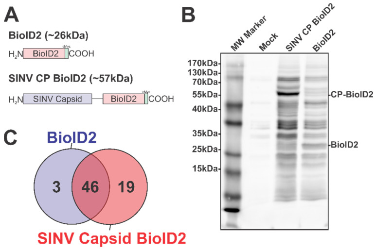
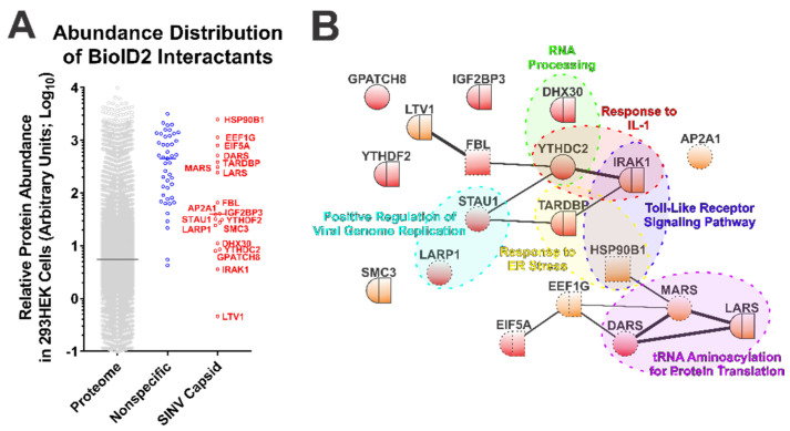
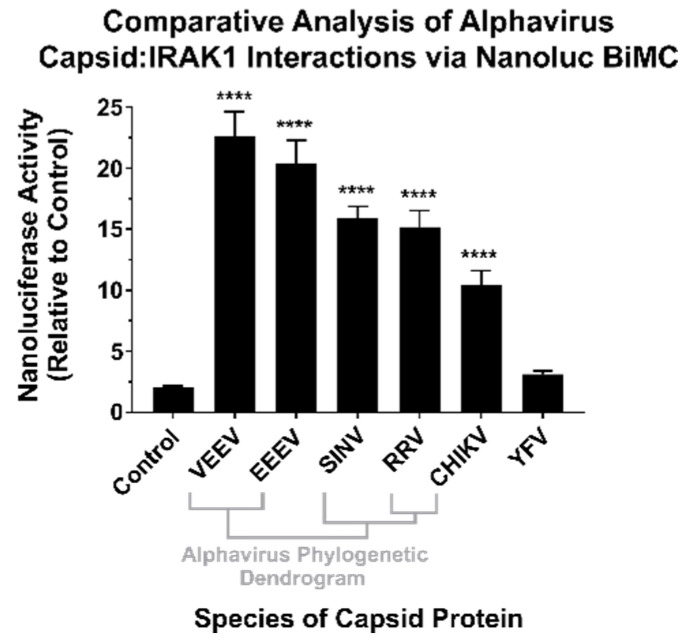
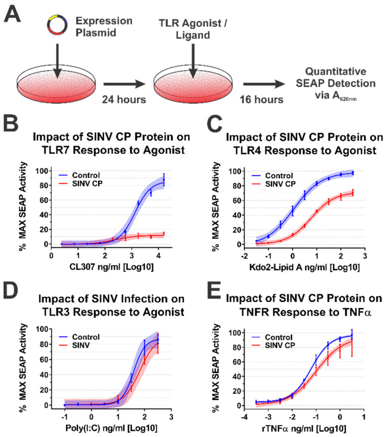
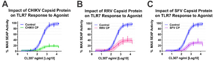
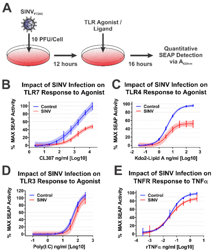
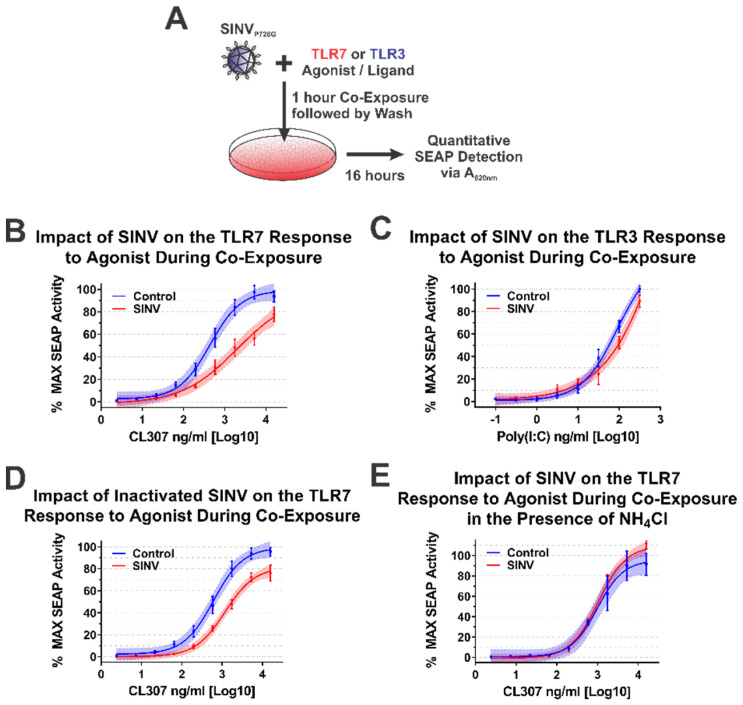
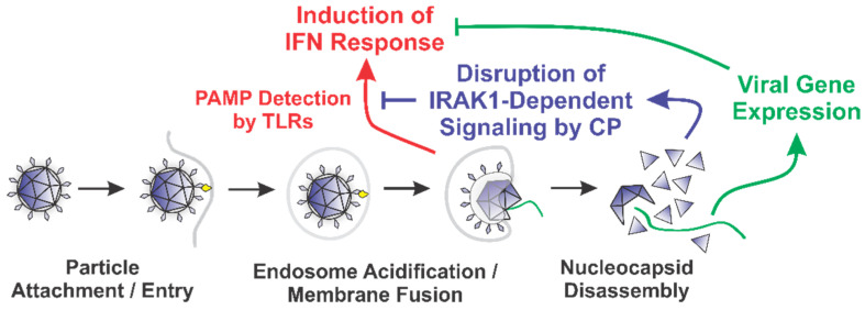
Alphaviruses are arthropod-borne RNA viruses which can cause either mild to severe febrile arthritis which may persist for months, or encephalitis which can lead to death or lifelong cognitive impairments. The non-assembly molecular role(s), functions, and protein-protein interactions of the alphavirus capsid proteins have been largely overlooked. Here we detail the use of a BioID2 biotin ligase system to identify the protein-protein interactions of the Sindbis virus capsid protein. These efforts led to the discovery of a series of novel host-pathogen interactions, including the identification of an interaction between the alphaviral capsid protein and the host IRAK1 protein. Importantly, this capsid-IRAK1 interaction is conserved across multiple alphavirus species, including arthritogenic alphaviruses SINV, Ross River virus, and Chikungunya virus; and encephalitic alphaviruses Eastern Equine Encephalitis virus, and Venezuelan Equine Encephalitis virus. The impact of the capsid-IRAK1 interaction was evaluated using a robust set of cellular model systems, leading to the realization that the alphaviral capsid protein specifically inhibits IRAK1-dependent signaling. This inhibition represents a means by which alphaviruses may evade innate immune detection and activation prior to viral gene expression. Altogether, these data identify novel capsid protein-protein interactions, establish the capsid-IRAK1 interaction as a common alphavirus host-pathogen interface, and delineate the molecular consequences of the capsid-IRAK1 interaction on IRAK1-dependent signaling.

Keywords: IRAK1; alphavirus; capsid; toll like receptors (TLR).


Conflict of interest statement

The authors declare no conflict of interest. The funding agencies had no role in the study design, data collection and analysis, decision to publish, or the preparation of the manuscript.

Figures

Figure 1
Figure 1
The Identification of the Host–Pathogen Interactions of the SINV Capsid Protein. (A) A diagram of the BioID2 fusion proteins expressed in 293HEK cells via plasmid transfection. Individual domains are labeled above. The line in the SINV CP BioID2 construct represents a poly-glycine linker, and the green box represents a cMyc tag. (B) A representative blot of 293HEK cell lysates after the BioID2 approach. Briefly, transfected or control transfected cells were cultured in the presence of excess biotin prior to the generation of whole-cell lysates. Equal protein amounts were resolved using SDS-PAGE, and subsequently probed for protein biotinylation using streptavidin-HRP. (C) A Venn diagram of the host proteins identified by mass spectrometry after the BioID2 approach designated the host factors as either non-specific or specific to either BioID2 transfection/purification.
Figure 2
Figure 2
Ontological Analysis of the SINV CP–Protein Interactants Reveals Novel Host–Pathogen Interfaces. (A) Comparative analysis of arbitrary protein abundance of the host proteome (293HEK), and the non-specific and CP-specific interactants identified via the BioID2 approach. The lines on the graph represent the median abundance within the given data set, and the CP-specific interactants are indicated next to their corresponding data point. (B) A STRINGs interaction network map of the CP-specific interactants. The color and styling of the individual nodes indicates the properties of the corresponding protein as determined by ontological categorization: round = cytoplasmic localization; square = nuclear localization; round/square = shuttling protein, or found in both compartments; red = RNA-associated protein; dashed outline = membrane associated. The weight of the linear connections between the individual nodes is indicative of the relative strength/confidence of the interaction. Molecular function ontological groups, as described in depth in the text, are highlighted in a color-coded manner.
Figure 3
Figure 3
The CP–IRAK1 Interaction Is Genuine, and Widely Conserved across the Genus Alphavirus. The interaction of the alphaviral CP proteins with the host IRAK1 protein was assessed using Nanoluc-based BiMolecular Complementation (BiMC). Briefly, 293HEK cells were co-transfected with expression plasmids encoding the human IRAK1 protein and one of the indicated CP proteins or the BioID2 protein fused to complementary fragments of Nanoluc. Forty-eight hours post transfection the cells were assayed using the NanoGlo live-cell assay system, and the luminescence was detected using a plate reader. The luminescent intensity of the CP–IRAK1 BiMC conditions was compared relative to those of control reactions lacking an interacting pair of Nanoluc fragments. The quantitative data shown are the mean of at least five biological replicates, with the error bar representing the standard deviation of the means. Statistical significance relative to the control reactions, with a p-value of < 0.0001 = ****, was determined by one-way ANOVA analysis. Below the X axis is a phylogenetic dendrogram of alphavirus CP amino acid relatedness.
Figure 4
Figure 4
The SINV Capsid Protein Inhibits IRAK1-Dependent Signaling in a Specific Manner. (A) A diagram of the experimental approach used to test the capacity of the SINV CP protein to inhibit IRAK1-dependent signaling in a specific manner. Comparison of the curves in each panel reveals the impact of SINV CP protein expression on (B) TLR7 activation by CL307, (C) TLR4 activation by Kdo2-lipid A, (D) TLR3 activation by poly(I:C), and (E) TNFR activation by rTNFα. In all graphs, cells receiving control transfections prior to agonist treatment are represented by blue lines and data points, and those receiving the SINV CP protein expression plasmid are represented by red lines and data points. All quantitative data shown are the minimum of six independent biological replicates conducted over several days with at least two independent plasmid preparations. Quantitative data shown are the means of the biological replicates, and the error bars represent the standard deviation of the means. The connecting line represents a non-linear regression of the underlying data, and the shaded region indicates the 95% confidence interval of the non-linear regression. Thus, data points where the shaded regions do not intersect are statistically significant by at least a p-value of <0.05, as determined by ANOVA analysis.
Figure 5
Figure 5
Old World Alphavirus Capsid Proteins Inhibit IRAK1-Dependent TLR7 Signaling. Comparison of the curves in each panel above reveals the impact of the Old World Alphaviral CP proteins expression on TLR7 activation by CL307. Specifically, the impact of ectopic expression of (A) CHIKV CP protein, (B) RRV CP protein, and (C) SFV CP proteins were assayed identically to that described for Figure 4. Quantitative data shown are the means of the biological replicates, and the error bars represent the standard deviation of the means. The connecting line represents a non-linear regression of the underlying data, and the shaded region indicates the 95% confidence interval of the non-linear regression. Thus, data points where the shaded regions do not intersect are statistically significant by at least a p-value of < 0.05, as determined by ANOVA analysis.
Figure 6
Figure 6
The SINV Infection Inhibits IRAK1-Dependent Signaling. (A) A diagram of the experimental approach used to test the capacity of SINVP726G to inhibit IRAK1-dependent signaling in a specific manner during infection. Comparison of the curves in each panel reveals the impact of SINV infection on (B) TLR7 activation by CL307, (C) TLR4 activation by Kdo2-lipid A, (D) TLR3 activation by poly(I:C), and (E) TNFR activation by rTNFα. In all graphs, cells mock infected prior to agonist treatment are represented by blue lines and data points, and those receiving infectious SINVP726G represented by red lines and data points. All quantitative data shown are the minimum of six independent biological replicates conducted over several days with at least two independent SINV preparations. Quantitative data shown are the means of the biological replicates, and the error bars represent the standard deviation of the means. The connecting line represents a non-linear regression of the underlying data, and the shaded region indicates the 95% confidence interval of the non-linear regression. Thus, data points where the shaded regions do not intersect are statistically significant by at least a p-value of < 0.05, as determined by ANOVA analysis.
Figure 7
Figure 7
The SINV Capsid Protein Delivered by Incoming Infectious and Non-Infectious Particles Is Sufficient to Inhibit IRAK1-Dependent TLR Signaling. (A) A representative diagram of the co-exposure systems used to assess the impact of the incoming SINV CP proteins derived from particles. Specific differences between the experimental designs are noted in the title of each graph. Comparison of the curves in each panel reveals the impact of the CP–IRAK1 interaction on agonist co-exposure during (B) delivery of the SINV CP protein from infectious particles in the presence of the TLR7 agonist CL307, (C) delivery of the SINV CP protein from infectious particles in the presence of the TLR3 agonist poly(I:C), (D) delivery of the SINV CP protein from UV inactivated particles in the presence of the TLR7 agonist CL307, and (E) the effect of viral entry inhibitors on the sensing of CL307 by TLR7. In all graphs, mock infected cells are represented by blue lines and data points, and those receiving SINV viral particles are represented by red lines and data points. All quantitative data shown are the minimum of six independent biological replicates conducted over several days with at least two independent SINV preparations. Quantitative data shown are the means of the biological replicates, and the error bars represent the standard deviation of the means. The connecting line represents a non-linear regression of the underlying data, and the shaded region indicates the 95% confidence interval of the non-linear regression. Thus, data points where the shaded regions do not intersect are statistically significant by at least a p-value of <0.05, as determined by ANOVA analysis.
Figure 8
Figure 8
A Diagram of the Roles of the Nucleocapsid Components Early during Infection.

Similar articles

Cited by

References

    1. Strauss J.H., Strauss E.G. The alphaviruses: Gene expression, replication, and evolution. Microbiol. Rev. 1994;58:491–562. doi: 10.1128/MR.58.3.491-562.1994. - DOI - PMC - PubMed
    1. Gao X., Nasci R., Liang G. The Neglected Arboviral Infections in Mainland China. PLoS Negl. Trop. Dis. 2010;4:e624. doi: 10.1371/journal.pntd.0000624. - DOI - PMC - PubMed
    1. Turunen M., Kuusisto P., E Uggeldahl P., Toivanen A. Pogosta disease: Clinical observations during an outbreak in the province of North Karelia, Finland. Rheumatology. 1998;37:1177–1180. doi: 10.1093/rheumatology/37.11.1177. - DOI - PubMed
    1. Adouchief S., Smura T., Sane J., Vapalahti O., Kurkela S. Sindbis virus as a human pathogen-epidemiology, clinical picture and pathogenesis. Rev. Med. Virol. 2016;26:221–241. doi: 10.1002/rmv.1876. - DOI - PubMed
    1. Hesse R.R., Weaver S.C., De Siger J., Medina G., Salas R.A. Emergence of a new epidemic/epizootic Venezuelan equine encephalitis virus in South America. Proc. Natl. Acad. Sci. USA. 1995;92:5278–5281. doi: 10.1073/pnas.92.12.5278. - DOI - PMC - PubMed

Publication types

MeSH terms

LinkOut - more resources