. 2021 Feb 27;22(5):2382.
doi: 10.3390/ijms22052382.

Mitochondrial Transfer Improves Cardiomyocyte Bioenergetics and Viability in Male Rats Exposed to Pregestational Diabetes

Affiliations

Mitochondrial Transfer Improves Cardiomyocyte Bioenergetics and Viability in Male Rats Exposed to Pregestational Diabetes

Eli J Louwagie et al. Int J Mol Sci. .

Abstract

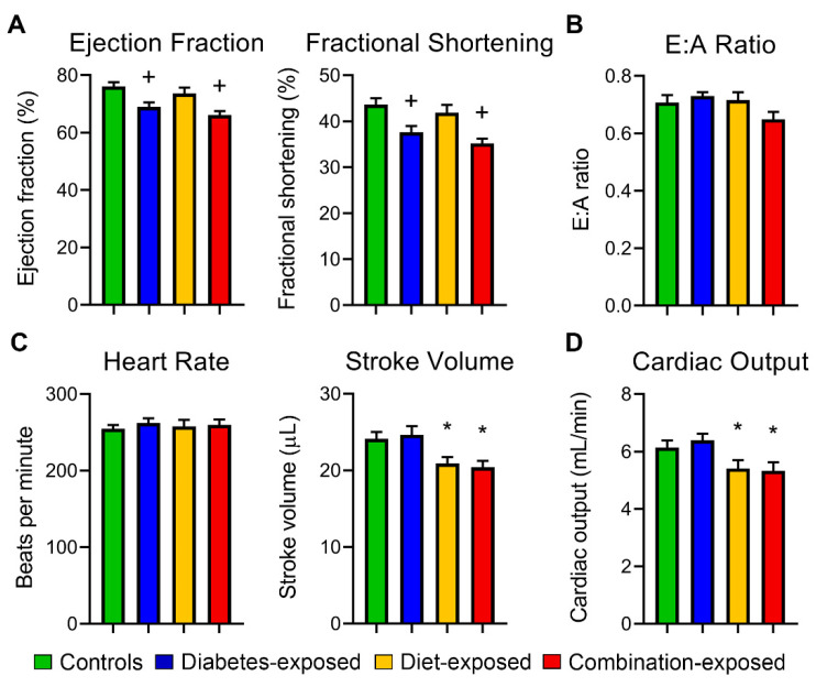
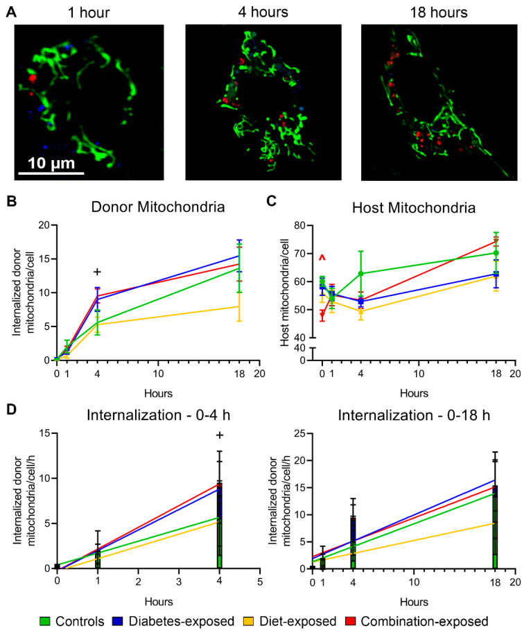
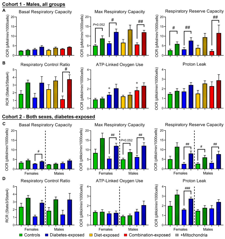
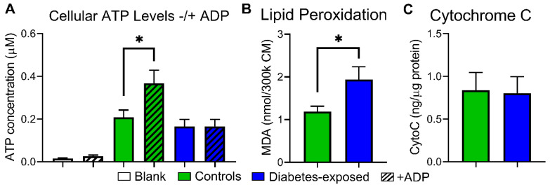
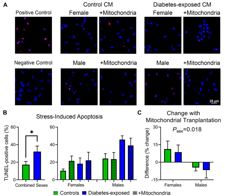
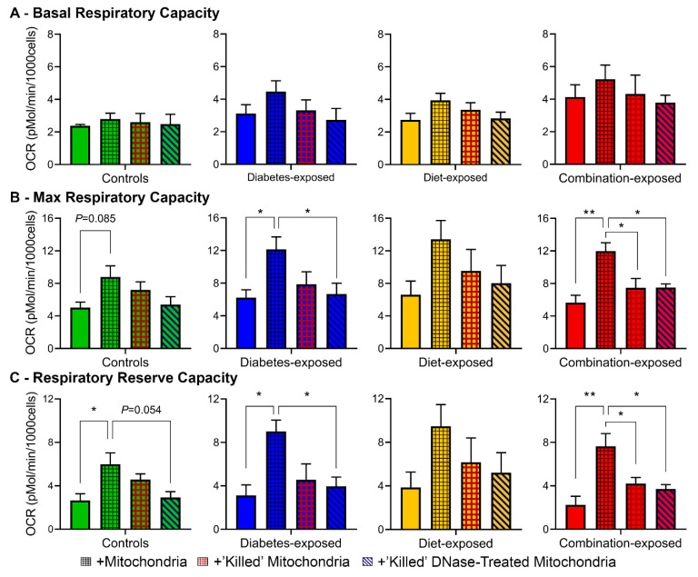
Offspring born to diabetic or obese mothers have a higher lifetime risk of heart disease. Previously, we found that rat offspring exposed to late-gestational diabetes mellitus (LGDM) and maternal high-fat (HF) diet develop mitochondrial dysfunction, impaired cardiomyocyte bioenergetics, and cardiac dysfunction at birth and again during aging. Here, we compared echocardiography, cardiomyocyte bioenergetics, oxidative damage, and mitochondria-mediated cell death among control, pregestational diabetes mellitus (PGDM)-exposed, HF-diet-exposed, and combination-exposed newborn offspring. We hypothesized that PGDM exposure, similar to LGDM, causes mitochondrial dysfunction to play a central, pathogenic role in neonatal cardiomyopathy. We found that PGDM-exposed offspring, similar to LGDM-exposed offspring, have cardiac dysfunction at birth, but their isolated cardiomyocytes have seemingly less bioenergetics impairment. This finding was due to confounding by impaired viability related to poorer ATP generation, more lipid peroxidation, and faster apoptosis under metabolic stress. To mechanistically isolate and test the role of mitochondria, we transferred mitochondria from normal rat myocardium to control and exposed neonatal rat cardiomyocytes. As expected, transfer provides a respiratory boost to cardiomyocytes from all groups. They also reduce apoptosis in PGDM-exposed males, but not in females. Findings highlight sex-specific differences in mitochondria-mediated mechanisms of developmentally programmed heart disease and underscore potential caveats of therapeutic mitochondrial transfer.

Keywords: developmentally programmed heart disease; diabetic pregnancy; mitochondria; mitochondrial transfer.


Conflict of interest statement

The authors declare no competing interest.

Figures

Figure 1
Figure 1
Diabetes- and high-fat (HF) diet-exposed newborn males had significant cardiac dysfunction at birth. (A) Systolic function, (B) diastolic function, (C) heart rate and stroke volume were determined in P1 offspring by echocardiography. (D) Cardiac output was calculated from (C). N = 12–19 offspring/group. Significant differences (p ≤ 0.05): + diabetes or * diet effect by 2-way NOVA.
Figure 2
Figure 2
Donor mitochondria are internalized by cardiomyocytes at faster rates with diabetes exposure. (A) Representative confocal live-cell images of MitoTracker Green- and LysoTracker Blue-stained cardiomyocytes co-incubated with pHrodo Red-stained donor mitochondria. Numbers of internalized donor mitochondria (B) and host mitochondria (C) over time. (D) Rates of donor mitochondria internalization in the first four hours of co-incubation (left) and across 18 h (right). Data represent mean ± SEM. N = 5–6 males/group. Significant differences (p ≤ 0.05): + diabetes effect by 2-way ANOVA, ^ group effect by 1-way ANOVA and Dunnett post hoc test when interaction was present by 2-way ANOVA.
Figure 3
Figure 3
PGDM-exposed males, but not females, use more oxygen for ATP production, and while mitochondrial transfer may benefit males, it may put females at greater risk of ROS production. (A) Basal, FCCP-stimulated maximal, and reserve respiratory capacities and (B) respiratory control ratios, oxygen consumed in ATP production, and proton leak of male cardiomyocytes (CM) determined by mitochondrial stress test supplemented with mitochondrial transfer. (C,D) Respiratory parameters from PGDM-exposed CM of both sexes. N = 4–6 per group. Data represent mean ± SEM. P ≤ 0.05: + diabetes effect by 2-way ANOVA, # mitochondrial effect by 1-way ANOVA. ## P < 0.01, ### P < 0.001.
Figure 4
Figure 4
PGDM-exposed cardiomyocytes suffer from impaired phosphorylation and increased lipid peroxidation. (A) Cellular ATP levels with and without addition of ADP. Left two columns show media (negative control). (B) Malondialdehyde (MDA) was measured as a surrogate marker for lipid peroxidation. (C) Cytosolic cytochrome C levels at baseline, i.e., not under stress. N = 8–10 per group (sexes combined). Data represent mean ± SEM. * P ≤ 0.05 by 1-way ANOVA (A) or T-test (B).
Figure 5
Figure 5
Females and PGDM-exposed cells have stronger mitochondrial membrane potential (MMP), but they lose it faster following metabolic stress. (A) Representative images of CM stained with MitoTracker Green, Hoechst, and MitoTracker Red. Once treated with FCCP, mitochondria lose their MMP and trigger cell death through intrinsic apoptosis. (B) MMP loss was analyzed before and following FCCP-induced stress by linear regression analysis. (C) Baseline MMP and (D) rate of MMP loss over 25 min of FCCP-induced stress. N = 4–5 per sex per group. Data represent mean ± SEM. P ≤ 0.05: + diabetes, # mitochondrial, or δ sex-specific effect (only lower sex marked) by 1-way ANOVA.
Figure 6
Figure 6
PGDM-exposed cardiomyocytes of both sexes are at greater risk of stress-induced apoptosis; mitochondrial transfer lowers this risk in males but worsens it in females. (A) Representative CM labeled with 4′,6-diamidino-2-phenylindole (DAPI) and transferase dUTP nick end labeling (TUNEL) for apoptotic cell death following FCCP Challenge. DNase treatment was used as a positive control. TUNEL reaction without enzyme was used as a negative control. (B) Percentage of TUNEL-positive cells following FCCP Challenge. Males have higher numbers of TUNEL-positive cells regardless of group (P = 0.005 by 1-way ANOVA; not demarcated). (C) Difference in TUNEL-positive cells with mitochondrial transfer. N = 4–5 per sex per group using 138 ± 10 cells per plate from 7–10 systematically imaged fields. Data represent mean ± SEM. * P ≤ 0.05 by 1-way ANOVA.
Figure 7
Figure 7
MtDNA plays a role in respiratory boost of mitochondrial transfer. (A) Basal, (B) FCCP-stimulated maximal, and (C) reserve respiratory capacities of CM treated with sham (solid), living–respiring (black hashing), “killed” (red hashing), and “killed”+DNase-treated mitochondria (diagonal). N = 4–6 per group. Data represent mean ± SEM. P ≤ 0.05: # mitochondrial effect by 1-way ANOVA. ** P < 0.01.

Similar articles

Cited by

References

    1. Riskin A., Garcia-Prats J.A. Infants of women with diabetes. In: Weisman L.E., Wolfsdorf J.I., Kim M.S., editors. UpToDate. Wolters Kluwer; Online: 2020.
    1. Depla A.L., de Wit L., Steenhuis T.J., Slieker M.G., Voormolen D.N., Scheffer P.G., de Heus R., van Rijn B.B., Bekker M.N. Effects of maternal diabetes on fetal heart function at echocardiography: Systematic review and meta-analysis. Ultrasound Obstet. Gynecol. 2020 doi: 10.1002/uog.22163. - DOI - PMC - PubMed
    1. Ullmo S., Vial Y., Di Bernardo S., Roth-Kleiner M., Mivelaz Y., Sekarski N., Ruiz J., Meijboom E.J. Pathologic ventricular hypertrophy in the offspring of diabetic mothers: A retrospective study. Eur. Heart J. 2007;28:1319–1325. doi: 10.1093/eurheartj/ehl416. - DOI - PubMed
    1. Nyrnes S.A., Garnæs K.K., Salvesen Ø., Timilsina A.S., Moholdt T., Ingul C.B. Cardiac function in newborns of obese women and the effect of exercise during pregnancy. A randomized controlled trial. PLoS ONE. 2018;13:e0197334. doi: 10.1371/journal.pone.0197334. - DOI - PMC - PubMed
    1. Stuart A., Amer-Wåhlin I., Persson J., Källen K. Long-term cardiovascular risk in relation to birth weight and exposure to maternal diabetes mellitus. Int. J. Cardiol. 2013;168:2653–2657. doi: 10.1016/j.ijcard.2013.03.032. - DOI - PubMed

MeSH terms