. 2021 Feb 27;10(3):361.
doi: 10.3390/antiox10030361.

Selenium-Binding Protein 1 (SELENBP1) Supports Hydrogen Sulfide Biosynthesis and Adipogenesis

Affiliations

Selenium-Binding Protein 1 (SELENBP1) Supports Hydrogen Sulfide Biosynthesis and Adipogenesis

Elisa B Randi et al. Antioxidants (Basel). .

Abstract

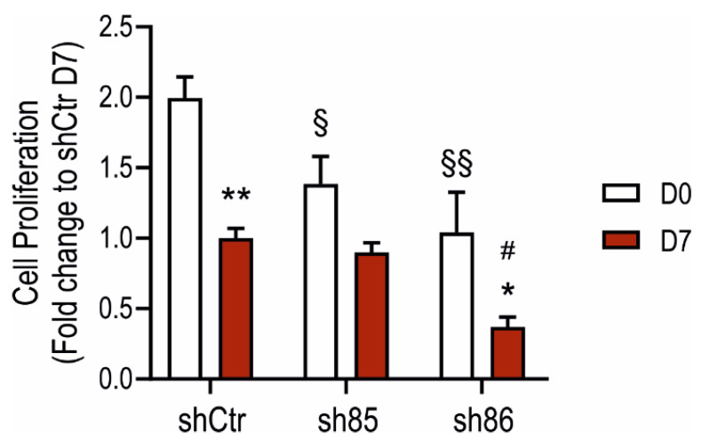
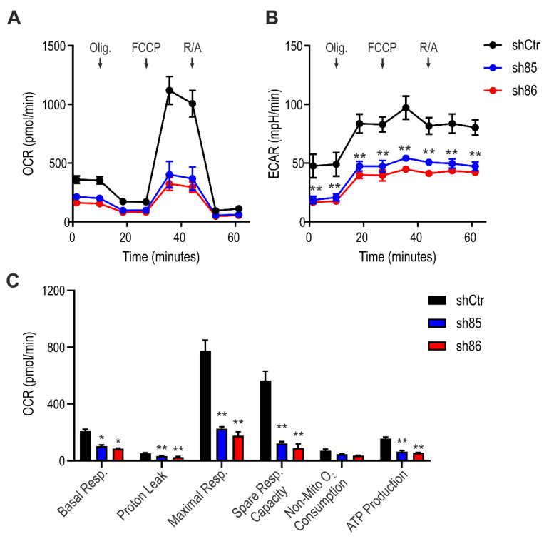
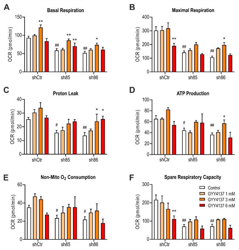
Hydrogen sulfide (H2S), a mammalian gasotransmitter, is involved in the regulation of a variety of fundamental processes including intracellular signaling, cellular bioenergetics, cell proliferation, and cell differentiation. Cystathionine g-lyase (CSE), cystathionine b-synthase (CBS), and 3-mercaptopyruvate sulfurtransferase (3-MST) are currently considered the three principal mammalian H2S-generating enzymes. However, recently, a fourth H2S-producing enzyme, selenium-binding-protein 1 (SELENBP1), has also been identified. The cellular regulatory role(s) of SELENBP1 are incompletely understood. The current study investigated whether SELENBP1 plays a role in the regulation of adipocyte differentiation in vitro. 3T3-L1 preadipocytes with or without SELENBP1 knock-down were subjected to differentiation-inducing conditions, and H2S production, cellular lipid accumulation, cell proliferation, and mitochondrial activity were quantified. Adipocyte differentiation was associated with an upregulation of H2S biosynthesis. SELENBP1 silencing decreased cellular H2S levels, suppressed the expression of the three "classical" H2S-producing enzymes (CBS, CSE, and 3-MST) and significantly suppressed adipocyte differentiation. Treatment of SELENBP1 knock-down cells with the H2S donor GYY4137 partially restored lipid accumulation, increased cellular H2S levels, and exerted a bell-shaped effect on cellular bioenergetics (enhancement at 1 and 3 mM, and inhibition at 6 mM). We conclude that SELENBP1 in adipocytes (1) contributes to H2S biosynthesis and (2) acts as an endogenous stimulator of adipocyte differentiation.

Keywords: fat; gasotransmitters, mitochondria, differentiation, metabolism; obesity.


Conflict of interest statement

The authors declare no conflict of interest.

Figures

Figure 1
Figure 1
Assessment of the adipogenesis process in wild-type 3T3-L1 and shSELENBP1 cells. (A) Schematic overview of the differentiation process in 3T3-L1 cells. (B) Representative immunoblot showing the expression of SELENBP1 and the adipocyte marker Adiponectin in two independent passages of wild-type cells before and after differentiation. (C) Representative immunoblot of SELENBP1 and adiponectin in stably transfected cells (shCtr and two independent shSELENBP1s, named sh85 and sh86), and corresponding densitometry analysis. β-Actin was used as loading control. D0: Day 0 (non-differentiated cells, i.e., pre-adipocytes); D7: Day 7 (mature (differentiated) adipocytes). DM: Differentiation Medium; MM: Maintenance Medium. ** p < 0.01 shows significant inhibition of SELENBP1 expression compared to control.
Figure 2
Figure 2
Effect of SELENBP1 knock-down on differentiation-associated lipid accumulation in 3T3-L1 cells. (A) Representative pictures of Oil Red O staining in shCtr and shSELENBP1 (sh85 and sh86 clones) cells before (Day 0) and after (Day 7) differentiation (scale bar: 200 µm). (B): numerical quantification of the findings. ** p < 0.01 indicates a significant difference between D7 and corresponding D0 values; ## p < 0.01 indicates a significant difference between shSELENBP1 (D7) and shCtr (D7).
Figure 3
Figure 3
Increases in cellular H2S content during adipogenesis: role of SELENBP1. AzMC-assessed quantification of cellular H2S levels before and after differentiation in shCtr and shSELENBP1 cells (sh85, sh86 clones). * p < 0.05 shows a significant difference between D7 and the corresponding D0; # p < 0.05 shows a significant difference between shSELENBP1 (D7) and shCtr (D7).
Figure 4
Figure 4
Effect of SELENBP1 silencing on adipocyte proliferation. Quantification of cell proliferation in shCtr and shSELENBP1 cells before and after differentiation. * p < 0.05 and ** p < 0.01 show significant differences between D7 and corresponding D0; # p < 0.05 shows a significant difference between shSELENBP1(D7) and shCtr(D7): § p < 0.05 and §§ p < 0.01 show significant differences between shCtr (D0) and shSELENBP1(D0).
Figure 5
Figure 5
Effect of SELENBP1 silencing on the XTT-converting activity of adipocytes during differentiation. XTT conversion was quantified in control and SELENBP1 knock-down cells before and after differentiation. * p < 0.05 shows significant difference between D7 and corresponding D0; # p < 0.05 shows significant difference between shSELENBP1 (D7) and shCtr (D7); § p < 0.05 shows significant difference between shCtr (D0) and shSELENBP1 (D0).
Figure 6
Figure 6
Effect of SELENBP1 silencing on mitochondrial respiration of mature adipocytes. (A) Oxygen consumption rate (OCR) and (B) extracellular acidification rate (ECAR) was measured in control and SELENBP1 knock-down cells after injection of oligomycin (Olig.), trifluoromethoxy carbonylcyanide phenylhydrazone (FCCP,) and Rotenone/antimycin A (AMA). (C) Analysis of different bioenergetics parameters obtained from OCR values. * p < 0.05 and ** p < 0.01 show significant differences between shSELENBP1 and shCtr.
Figure 7
Figure 7
Effect of GYY4137 on H2S-producing enzymes. (A) Schematic representation of GYY4137 treatment during the differentiation process. (B) Representative immunoblots of SELENBP1, CBS, CSE, and 3-MST in shCtr and ShSELENBP1 cells (clones sh85 and sh86), differentiated in the presence of the H2S donor GYY4137. (C) Quantification of the densitometric data. * p < 0.05 and ** p < 0.01 show significant differences between GYY4137-treated cells and the corresponding control.
Figure 8
Figure 8
Effect of the H2S donor GYY4137 on differentiation-associated lipid accumulation in shSELENBP1 knock-down cells. (A) quantification of Oil Red O staining in shCtr and shSELENBP1 (sh85, sh86 clones) treated with 1, 3, and 6 mM GYY4137. * p < 0.05 indicates a significant difference between GYY4137-treated and corresponding untreated values; # p < 0.05 indicates a significant difference between shSELENBP1 (untreated) and shCtr (untreated).
Figure 9
Figure 9
Increased cellular H2S content in SELENBP1 knock-down cells treated with GYY4137. AzMC-assessed quantification of cellular H2S levels in shCtr and shSELENBP1 cells (sh85, sh86 clones) treated with GYY4137 during the differentiation process. * p < 0.05 shows a significant difference between GYY4137-treated cells and the group control; ## p < 0.01 indicates a significant difference between shSELENBP1 (untreated) and shCtr (untreated).
Figure 10
Figure 10
Effect of GYY4137 on bioenergetics parameters in SELENBP1 knock-down cells. (A) Basal respiration, (B) maximal respiration, (C) proton leak, (D) ATP production, (E) non-mitochondrial oxygen consumption, and (F) spare respiratory capacity were calculated after extracellular flux analysis on GYY4137-treated shSELENBP1 cells. ** p < 0.01 shows a significant difference between GYY4137-treated cells and the group control; # p < 0.05 and ## p < 0.01 indicate significant differences between shSELENBP1 (untreated) and shCtr (untreated).

Similar articles

Cited by

References

    1. Szabo C. A timeline of hydrogen sulfide (H2S) research: From environmental toxin to biological mediator. Biochem. Pharmacol. 2018;149:5–19. doi: 10.1016/j.bcp.2017.09.010. - DOI - PMC - PubMed
    1. Aroca A., Gotor C., Bassham D.C., Romero L.C. Hydrogen sulfide: From a toxic molecule to a key molecule of cell life. Antioxidants. 2020;9:621. doi: 10.3390/antiox9070621. - DOI - PMC - PubMed
    1. Kimura H. Hydrogen sulfide as a neuromodulator. Mol. Neurobiol. 2002;26:13–19. doi: 10.1385/MN:26:1:013. - DOI - PubMed
    1. Szabo C. Hydrogen sulphide and its therapeutic potential. Nat. Rev. Drug Discov. 2007;6:917–935. doi: 10.1038/nrd2425. - DOI - PubMed
    1. Kimura H. Hydrogen sulfide: Its production, release and functions. Amino Acids. 2011;41:113–121. doi: 10.1007/s00726-010-0510-x. - DOI - PubMed

LinkOut - more resources