. 2021 Feb 27;11(3):596.
doi: 10.3390/nano11030596.

Calcium Silicate-Based Biocompatible Light-Curable Dental Material for Dental Pulpal Complex

Affiliations

Calcium Silicate-Based Biocompatible Light-Curable Dental Material for Dental Pulpal Complex

Sung-Min Park et al. Nanomaterials (Basel). .

Abstract

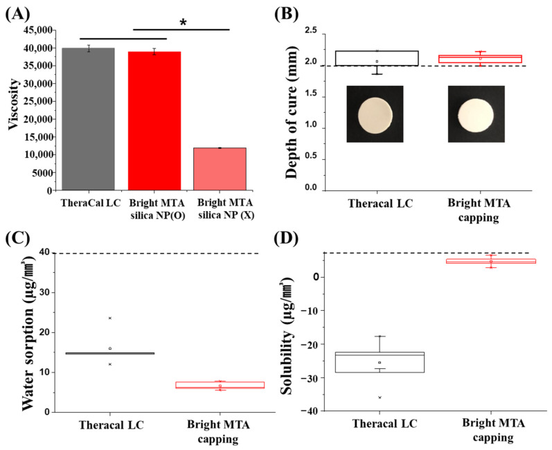
Dental caries causes tooth defects and clinical treatment is essential. To prevent further damage and protect healthy teeth, appropriate dental material is a need. However, the biocompatibility of dental material is needed to secure the oral environment. For this purpose, biocompatible materials were investigated for incorporated with dental capping material. Among them, nanomaterials are applied to dental materials to enhance their chemical, mechanical, and biological properties. This research aimed to study the physicochemical and mechanical properties and biocompatibility of a recently introduced light-curable mineral trioxide aggregate (MTA)-like material without bisphenol A-glycidyl methacrylate (Bis-GMA). To overcome the compromised mechanical properties in the absence of Bis-GMA, silica nanoparticles were synthesized and blended with a dental polymer for the formation of a nano-network. This material was compared with a conventional light-curable MTA-like material that contains Bis-GMA. Investigation of the physiochemical properties followed ISO 4049. Hydroxyl and calcium ion release from the materials was measured over 21 days. The Vickers hardness test and three-point flexural strength test were used to assess the mechanical properties. Specimens were immersed in solutions that mimicked human body plasma for seven days, and surface characteristics were analyzed. Biological properties were assessed by cytotoxicity and biomineralization tests. There was no significant difference between the tested materials with respect to overall physicochemical properties and released calcium ions. The newly produced material released more calcium ions on the third day, but 14 days later, the other material containing Bis-GMA released higher levels of calcium ions. The microhardness was reduced in a low pH environment, and differences between the specimens were observed. The flexural strength of the newly developed material was significantly higher, and different surface morphologies were detected. The recently produced extract showed higher cell viability at an extract concentration of 100%, while mineralization was clear at the conventional concentration of 25%. No significant changes in the physical properties between Bis-GMA incorporate material and nanoparticle incorporate materials.

Keywords: calcium silicate; light-curable MTA; nanoparticle; odontogenic differentiation; pulp regenerative dental materials.


Conflict of interest statement

The authors declare no conflicts of interest. The funders had no role in the design of the study; in the collection, analyses, or interpretation of data; in the writing of the manuscript, or in the decision to publish the results.

Figures

Figure 1
Figure 1
Physiochemical properties of TheraCal LC and bright MTA capping. (A) Viscosity comparison of TheraCal LC and bright MTA with silica nanoparticle (NP) or without (n = 5, at 24.7 °C p < 0.05). Asterisks (*) indicate statistical significance between specimen’s viscosity (p < 0.05). (B) Depth of cure (n = 5) of TheraCal and bright MTA capping was approximately 2.0 mm and 2.1 mm, respectively. According to ISO4049, opaque shade restorative materials are >2.0 mm. (C) Water absorption of TheraCal and bright MTA capping was 0.133 μg/mm3 and 0.066 μg/mm3 on average. According to ISO4049, at least four of the values obtained are ≤40 μg/mm3. (D) Water solubility (n = 5) of TheraCal and bright MTA capping was 0.025 μg/mm3, 0.004 μg/mm3 on average. According to ISO4049, at least four of the values obtained were ≤7.5 μg/mm3.
Figure 2
Figure 2
Inorganic amount and size distribution TheraCal LC and bright MTA. (A) Thermogravimetric analysis (TGA) results from 100 °C to 800 °C (n = 5) and (B) size distribution analysis after heat treatment at 800 °C (n = 5).
Figure 3
Figure 3
Ion release and pH changes from TheraCal LC and bright MTA capping over time. (A) The pH change was similar between TheraCal LC and bright MTA capping (n = 5). Asterisks (*) indicate statistical significance between bright MTA and TheraCal LC at the same pH (p < 0.05). The pH value fluctuated between pH 8 and 9 (p < 0.05, n = 5). (B) Calcium ions were similarly released for up to 10 days, with similar profile patterns between TheraCal LC and bright MTA capping, but after 10 days, TheraCal LC released more calcium ions than bright MTA capping (p < 0.05, n = 3). The ability of TheraCal LC and bright MTA capping to release calcium ions and alkalinize the surrounding fluids was correlated to the formation of calcium hydroxide Ca(OH)2.
Figure 4
Figure 4
Mechano-physical properties of nanoparticle-incorporated, light-curable MTA. (A) Vickers hardness measured in five spots per specimen and averaged (n = 10). Asterisks (*) indicate statistical significance between bright MTA and TheraCal LC at the same pH (p < 0.05). Different letters indicate statistical significance between bright MTA and TheraCal LC at the same pH (p < 0.05). This test attempted to simulate the actual clinical environment during inflammation. (B) Three-point flexural strength (n = 10). There was a statistical difference between bright MTA and TheraCal LC. The (#) symbol and asterisks (*) indicate bright MTA capping and TheraCal LC’s statistical significance based on the lowest value of micro-hardness (* p < 0.05, ** p < 0.01) (pH = 4). (C) Surface morphology of bright MTA and TheraCal LC immersed in SBF solution for seven days, as visualized by scanning electron microscopy (×10,000) (scale bar = 1 μm). The SBF solution can mimic the main features of blood serum; thus, using the SBF solution can test bioactivity. (D) XRD analysis of specimens immersed in SBF for seven days.
Figure 5
Figure 5
Cell viability and odontoblastic differentiation of nanoparticle-incorporated, light-curable MTA (A) Cell viability at various concentrations of extracts from TheraCal LC and bright MTA capping (n = 3, p < 0.05). Asterisks (*) indicate statistical significance between bright MTA and TheraCal LC’s cell viability based on the 0% of extract culture media. (#) the symbol indicates statistical significance between bright MTA and TheraCal LC’s cell viability at 100% extract culture media result. (B) Live and dead cells exposed to 25%, 50%, and 100% concentrations of extracts from bright MTA capping and TheraCal LC. Live (green) and dead (red) cells were observed by fluorescence microscopy (scale bar = 100 μm). Washing steps were performed before live and dead staining to ensure that only intact live cells remained on the plate (scale bar = 350 μm). (C) Odontoblastic differentiation under the use of Bright MTA and TheraCal LC was evaluated with alkaline phosphatase (ALP) activity (14 days) and calcium deposition (21 days) evaluated by alizarin red S (ARS) staining. ALP staining after 14 days revealed that 25% concentrations of extracts from both Bright MTA and TheraCal LC deposited a larger amount of phosphate than 50% concentrations. ARS staining after 21 days indicated a large amount of deposition in the 25% concentrations of extracts from both Bright MTA and TheraCal LC.

References

    1. Camilleri J., Laurent P., About I. Hydration of biodentine, theracal lc, and a prototype tricalcium silicate–based dentin replacement material after pulp capping in entire tooth cultures. J. Endod. 2014;40:1846–1854. doi: 10.1016/j.joen.2014.06.018. - DOI - PubMed
    1. Jo S.B., Kim H.K., Lee H.N., Kim Y.-J., Patel K.D., Campbell Knowles J., Lee J.-H., Song M. Physical properties and biofunctionalities of bioactive root canal sealers in vitro. Nanomaterials. 2020;10:1750. doi: 10.3390/nano10091750. - DOI - PMC - PubMed
    1. Mozayeni M.A., Milani A.S., Marvasti L.A., Asgary S. Cytotoxicity of calcium enriched mixture cement compared with mineral trioxide aggregate and intermediate restorative material. Aust. Endod. J. 2012;38:70–75. doi: 10.1111/j.1747-4477.2010.00269.x. - DOI - PubMed
    1. Poggio C., Lombardini M., Colombo M., Beltrami R., Rindi S. Solubility and pH of direct pulp capping materials: A comparative study. J. Appl. Biomater. Funct. Mater. 2015;13:181–185. doi: 10.5301/jabfm.5000230. - DOI - PubMed
    1. Gandolfi M., Siboni F., Prati C. Chemical–physical properties of TheraCal, a novel light-curable MTA-like material for pulp capping. Int. Endod. J. 2012;45:571–579. doi: 10.1111/j.1365-2591.2012.02013.x. - DOI - PubMed

LinkOut - more resources