

The Role of Autophagy in Eye Diseases
- PMID: 33673657
- PMCID: PMC7997177
- DOI: 10.3390/life11030189
The Role of Autophagy in Eye Diseases
Abstract
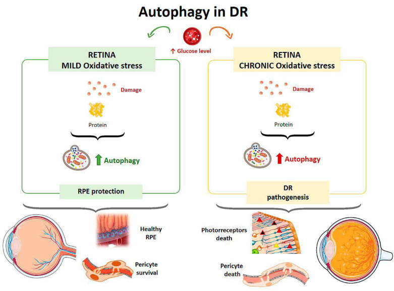
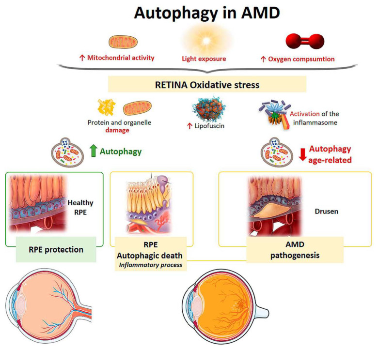
Autophagy is a catabolic process that ensures homeostasis in the cells of our organism. It plays a crucial role in protecting eye cells against oxidative damage and external stress factors. Ocular pathologies of high incidence, such as age-related macular degeneration, cataracts, glaucoma, and diabetic retinopathy are of multifactorial origin and are associated with genetic, environmental factors, age, and oxidative stress, among others; the latter factor is one of the most influential in ocular diseases, directly affecting the processes of autophagy activity. Alteration of the normal functioning of autophagy processes can interrupt organelle turnover, leading to the accumulation of cellular debris and causing physiological dysfunction of the eye. The aim of this study is to review research on the role of autophagy processes in the main ocular pathologies, which have a high incidence and result in high costs for the health system. Considering the role of autophagy processes in cell homeostasis and cell viability, the control and modulation of autophagy processes in ocular pathologies could constitute a new therapeutic approach.
Keywords: AMD; autophagy; cataract; diabetic retinopathy; glaucoma; ocular pathology.
Conflict of interest statement
The authors declare no conflict of interest.
Figures
Similar articles
-
The Evolving Functions of Autophagy in Ocular Health: A Double-edged Sword.Int J Biol Sci. 2016 Oct 25;12(11):1332-1340. doi: 10.7150/ijbs.16245. eCollection 2016. Int J Biol Sci. 2016. PMID: 27877085 Free PMC article. Review.
-
The role and therapeutic potential of melatonin in age-related ocular diseases.J Pineal Res. 2017 Sep;63(2). doi: 10.1111/jpi.12430. Epub 2017 Jul 18. J Pineal Res. 2017. PMID: 28658514 Review.
-
The interplay of autophagy and oxidative stress in the pathogenesis and therapy of retinal degenerative diseases.Cell Biosci. 2022 Jan 3;12(1):1. doi: 10.1186/s13578-021-00736-9. Cell Biosci. 2022. PMID: 34980273 Free PMC article. Review.
-
Autophagy and Age-Related Eye Diseases.Biomed Res Int. 2019 Dec 14;2019:5763658. doi: 10.1155/2019/5763658. eCollection 2019. Biomed Res Int. 2019. PMID: 31950044 Free PMC article. Review.
-
Autophagy in the eye: Development, degeneration, and aging.Prog Retin Eye Res. 2016 Nov;55:206-245. doi: 10.1016/j.preteyeres.2016.08.001. Epub 2016 Aug 23. Prog Retin Eye Res. 2016. PMID: 27566190 Review.
Cited by
-
Pharmacological Strategies for Cataract Management: From Molecular Targets to Clinical Translation.Int J Mol Sci. 2025 Jun 13;26(12):5658. doi: 10.3390/ijms26125658. Int J Mol Sci. 2025. PMID: 40565122 Free PMC article. Review.
-
Gene and phenome-based analysis of the shared genetic architecture of eye diseases.Am J Hum Genet. 2025 Feb 6;112(2):318-331. doi: 10.1016/j.ajhg.2025.01.004. Epub 2025 Jan 28. Am J Hum Genet. 2025. PMID: 39879988 Free PMC article.
-
The Role of Autophagy in Chemical Proteasome Inhibition Model of Retinal Degeneration.Int J Mol Sci. 2021 Jul 6;22(14):7271. doi: 10.3390/ijms22147271. Int J Mol Sci. 2021. PMID: 34298888 Free PMC article.
-
Retinal Pigment Epithelium Under Oxidative Stress: Chaperoning Autophagy and Beyond.Int J Mol Sci. 2025 Jan 30;26(3):1193. doi: 10.3390/ijms26031193. Int J Mol Sci. 2025. PMID: 39940964 Free PMC article. Review.
-
Regulation of TGF-β2-induced epithelial-mesenchymal transition and autophagy in lens epithelial cells by the miR-492/NPM1 axis.Biomol Biomed. 2024 Sep 6;24(5):1273-1289. doi: 10.17305/bb.2024.10249. Biomol Biomed. 2024. PMID: 38662949 Free PMC article.
References
Publication types
Grants and funding
LinkOut - more resources
Full Text Sources
Other Literature Sources
