. 2021 Feb 27;19(3):132.
doi: 10.3390/md19030132.

Fucoxanthin Suppresses Osteoclastogenesis via Modulation of MAP Kinase and Nrf2 Signaling

Affiliations

Fucoxanthin Suppresses Osteoclastogenesis via Modulation of MAP Kinase and Nrf2 Signaling

You-Jung Ha et al. Mar Drugs. .

Abstract

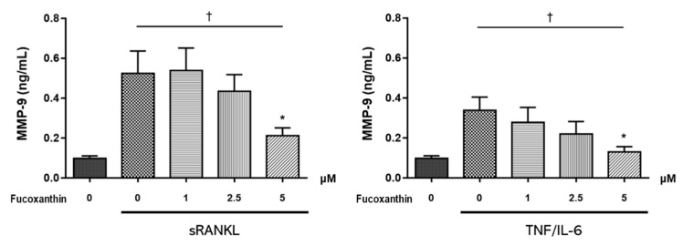
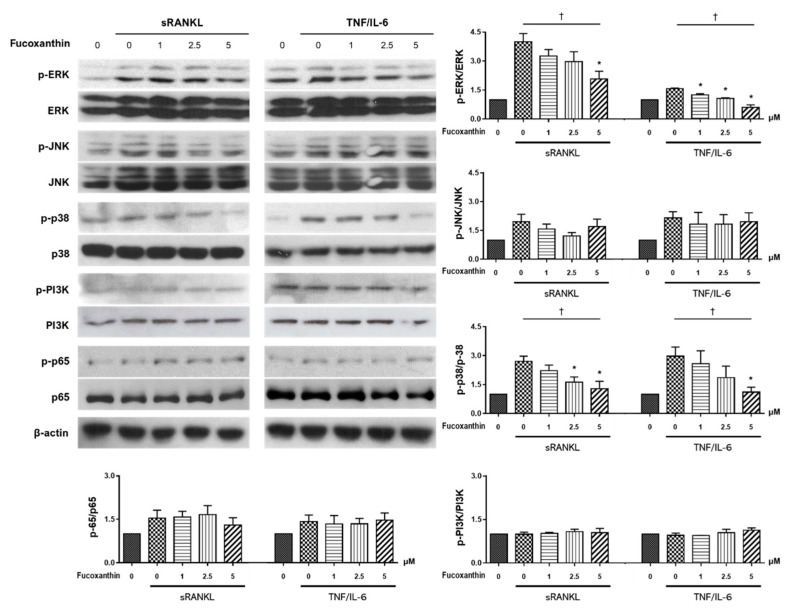
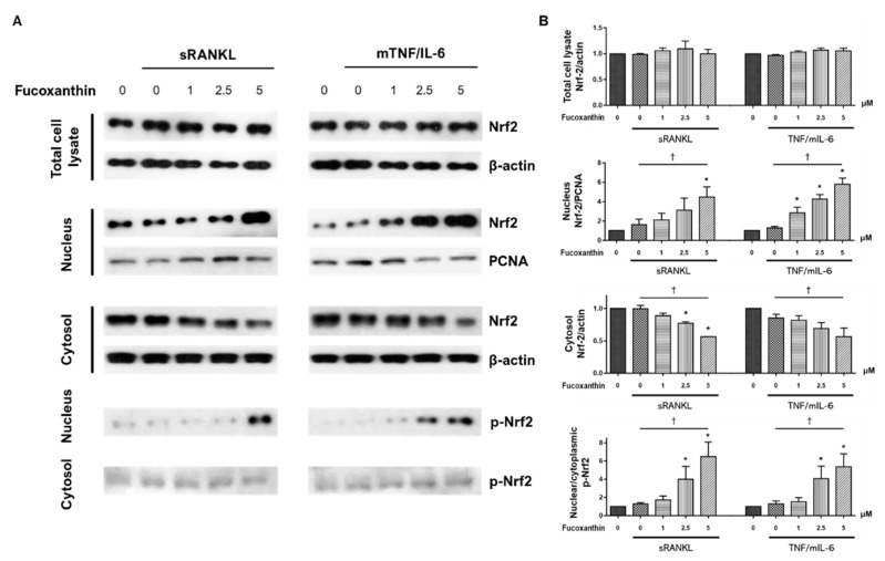
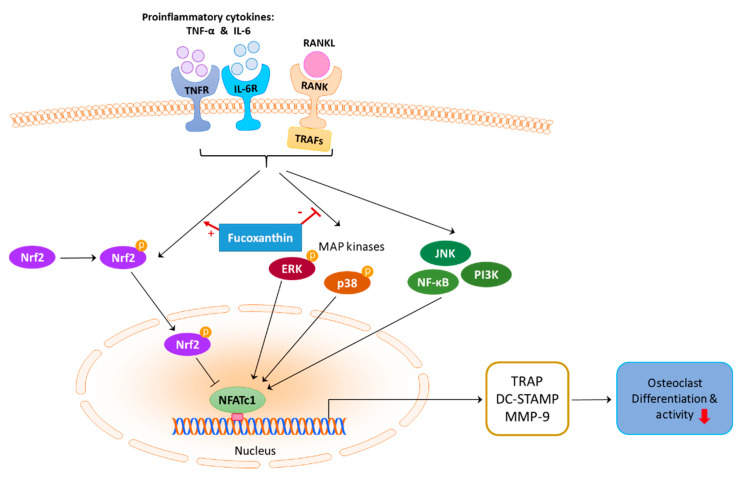
Fucoxanthin (FX), a natural carotenoid present in edible brown seaweed, is known for its therapeutic potential in various diseases, including bone disease. However, its underlying regulatory mechanisms in osteoclastogenesis remain unclear. In this study, we investigated the effect of FX on osteoclast differentiation and its regulatory signaling pathway. In vitro studies were performed using osteoclast-like RAW264.7 cells stimulated with the soluble receptor activator of nuclear factor-κB ligand or tumor necrosis factor-alpha/interleukin-6. FX treatment significantly inhibited osteoclast differentiation and bone resorption ability, and downregulated the expression of osteoclast-specific markers such as nuclear factor of activated T cells 1, dendritic cell-specific seven transmembrane protein, and matrix metallopeptidase 9. Intracellular signaling pathway analysis revealed that FX specifically decreased the activation of the extracellular signal-regulated kinase and p38 kinase, and increased the nuclear translocation of phosphonuclear factor erythroid 2-related factor 2 (Nrf2). Our results suggest that FX regulates the expression of mitogen-activated protein kinases and Nrf2. Therefore, FX is a potential therapeutic agent for osteoclast-related skeletal disorders including osteoporosis and rheumatoid arthritis.

Keywords: MAP kinase; Nrf2; brown seaweed; fucoxanthin; osteoclastogenesis.


Conflict of interest statement

The authors declare no conflict of interest.

Figures

Figure 1
Figure 1
Effect of fucoxanthin (FX) on viability of RAW264.7 cells. (A) Cells treated with different concentrations of FX, and cell viability was determined using 3-(4,5-dimethylthiazol-2-yl) -2,5-diphenyltetrazolium bromide (MTT) assay. Data are representative of six independent experiments and are expressed as mean ± standard error of mean (SEM); * p < 0.05 versus FX-untreated cells (0 μM). (B) Procaspase-3 and poly ADP ribose polymerase (PARP) expression remained uncleaved upon treatment with ≤10 μM FX, unlike in pemetrexed-treated cells of lung-cancer cell line NCI-H3122.
Figure 2
Figure 2
Soluble receptor activator of nuclear factor-κB (NF-κB) ligand (sRANKL)- or tumor necrosis factor (TNF)-α/interleukin (IL)-6-induced differentiation into osteoclast-like cells. (A) Representative microscopic images of tartrate-resistant acid phosphatase (TRAP) stained RAW264.7 cells (red arrows; original magnification, 100×). Blue box in bottom corner is a magnified photograph of the smaller boxed area (original magnification, 400×). (B,C) Number of TRAP-positive multinucleated cells differentiated from (B) RAW264.7 cells and (C) human CD14+ monocytes decreased upon treatment with FX in a dose-dependent manner. Data are representative of three independent experiments and are expressed as mean ± SEM; * p < 0.05 versus FX-untreated osteoclast-differentiated cells; p < 0.05 by Jonckheere–Terpstra test. FX, fucoxanthin.
Figure 3
Figure 3
Effect of FX on osteoclast activity. Statistical differences of resorption pit area and trends tests are presented in histograms. Data are representative of three independent experiments and are expressed as mean ± SEM. * p < 0.05 versus FX untreated cells; p < 0.05 by Jonckheere–Terpstra test.
Figure 4
Figure 4
Effect of FX treatment on expression of osteoclast-specific markers. (A) Nuclear factor of activated T cell 1 (NFATc1) protein expression and (B) dendritic-cell-specific transmembrane protein (DC-STAMP) mRNA expression in sRANKL- and TNF/IL-6-stimulated RAW264.7 cells decreased upon treatment with FX in a dose-dependent manner. Data are representative of three independent experiments and expressed as mean ± SEM; * p < 0.05 versus FX untreated cells; p < 0.05 by Jonckheere–Terpstra test.
Figure 5
Figure 5
Effect of FX treatment on MMP-9 levels in culture supernatant of osteoclast differentiated RAW264.7 cells. MMP-9 concentration significantly decreased upon treatment with 5 μM FX. Data are representative of three independent experiments and expressed as mean ± SEM; * p < 0.05 versus FX untreated cells; p < 0.05 by Jonckheere–Terpstra test.
Figure 6
Figure 6
Effect of FX treatment on signaling pathways during osteoclastogenesis. FX inhibited extracellular signal-regulated kinase (ERK) and p38 activation in both RANKL- and TNF-α/IL-6- stimulated conditions. However, c-Jun N-terminal kinase (JNK), phosphoinositide 3-kinase (PI3K), and NF-κB levels were not significantly altered. Data are representative of five independent experiments and expressed as mean ± SEM; * p < 0.05 versus FX untreated cells; p < 0.05 by Jonckheere–Terpstra test.
Figure 7
Figure 7
Effect of FX treatment on phosphorylated nuclear factor erythroid 2-related factor 2 (p-Nrf2) expression and nuclear localization of Nrf2 during osteoclast differentiation. Total cellular proteins were extracted from RAW264.7 cells and p-Nrf2 and Nrf2 expression were assessed by Western blotting. Cell lysates were fractionated into nuclear and cytosolic extracts, and identical experiments were performed. (A) Representative immunoblots and graphs for Nrf2 in total cell lysate, nucleus, and cytosol, and (B) nuclear/cytoplasmic p-Nrf2 from three independent experiments are shown. Data expressed as mean ± SEM; * p < 0.05 versus FX untreated cells; p < 0.05 by Jonckheere–Terpstra test.
Figure 8
Figure 8
Signaling pathways and effects of FX during osteoclastogenesis. Inhibitory effect of FX is mediated by blocking the activation of ERK and p38, promoting Nrf2 nuclear translocation and phosphorylation, and subsequently downregulating NFATc1. Nrf2 induction was previously reported to suppress NFATc1 transcriptional activity [57].

Similar articles

Cited by

References

    1. Walsh M.C., Kim N., Kadono Y., Rho J., Lee S.Y., Lorenzo J., Choi Y. Osteoimmunology: Interplay between the immune system and bone metabolism. Annu. Rev. Immunol. 2006;24:33–63. doi: 10.1146/annurev.immunol.24.021605.090646. - DOI - PubMed
    1. Boyle W.J., Simonet W.S., Lacey D.L. Osteoclast differentiation and activation. Nature. 2003;423:337–342. doi: 10.1038/nature01658. - DOI - PubMed
    1. Roodman G.D. Cell biology of the osteoclast. Exp. Hematol. 1999;27:1229–1241. doi: 10.1016/S0301-472X(99)00061-2. - DOI - PubMed
    1. Teitelbaum S.L. Bone resorption by osteoclasts. Science. 2000;289:1504–1508. doi: 10.1126/science.289.5484.1504. - DOI - PubMed
    1. Long C.L., Humphrey M.B. Osteoimmunology: The expanding role of immunoreceptors in osteoclasts and bone remodeling. BoneKEy Rep. 2012;1:59. doi: 10.1038/bonekey.2012.59. - DOI - PMC - PubMed

MeSH terms

LinkOut - more resources