Review
. 2021 Feb 27;13(5):1000.
doi: 10.3390/cancers13051000.

Subversion of Niche-Signalling Pathways in Colorectal Cancer: What Makes and Breaks the Intestinal Stem Cell

Affiliations
Review

Subversion of Niche-Signalling Pathways in Colorectal Cancer: What Makes and Breaks the Intestinal Stem Cell

Nathalie Sphyris et al. Cancers (Basel). .

Abstract

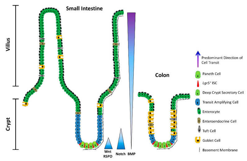
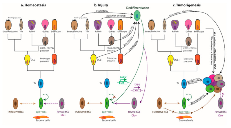
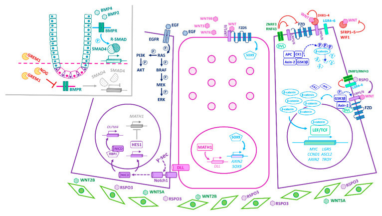
The intestinal epithelium fulfils pleiotropic functions in nutrient uptake, waste elimination, and immune surveillance while also forming a barrier against luminal toxins and gut-resident microbiota. Incessantly barraged by extraneous stresses, the intestine must continuously replenish its epithelial lining and regenerate the full gamut of specialized cell types that underpin its functions. Homeostatic remodelling is orchestrated by the intestinal stem cell (ISC) niche: a convergence of epithelial- and stromal-derived cues, which maintains ISCs in a multipotent state. Following demise of homeostatic ISCs post injury, plasticity is pervasive among multiple populations of reserve stem-like cells, lineage-committed progenitors, and/or fully differentiated cell types, all of which can contribute to regeneration and repair. Failure to restore the epithelial barrier risks seepage of toxic luminal contents, resulting in inflammation and likely predisposing to tumour formation. Here, we explore how homeostatic niche-signalling pathways are subverted in tumorigenesis, enabling ISCs to gain autonomy from niche restraints ("ISC emancipation") and transform into cancer stem cells capable of driving tumour initiation, progression, and therapy resistance. We further consider the implications of the pervasive plasticity of the intestinal epithelium for the trajectory of colorectal cancer, the emergence of distinct molecular subtypes, the propensity to metastasize, and the development of effective therapeutic strategies.

Keywords: BMP; Notch; Wnt; YAP; cancer stem cells (CSCs); colorectal cancer (CRC); consensus molecular subtypes (CMS); intestinal stem cell (ISC) niche; intestinal stem cells (ISCs); regeneration.


Conflict of interest statement

The authors declare no competing interests.

Figures

Figure 1
Figure 1
The architecture of the small intestine and the colon. Schematic depicting a longitudinal section of the intestinal mucosa. The mucosa of the small intestine extends finger-like projections (villi) into the gut lumen, which provide an increased surface area for optimal nutrient absorption. The villi are populated by mature, differentiated absorptive and secretory cell types, including absorptive enterocytes, hormone- and neurotransmitter-secreting enteroendocrine cells, mucus-secreting goblet cells, tuft cells, and microfold (M) cells (not shown). The mucosa surrounding the villi forms tubular invaginations into the lamina propria, called crypts, which serve as a protected reservoir of stem and progenitor cell populations. Notably, the epithelium of the colon is devoid of villi, with the crypts opening onto a flat mucosal surface, reflecting its role in waste compaction. To support homeostatic turnover, ISCs self-renew and give rise to short-lived transit-amplifying (TA) cells, which in turn beget lineage-restricted progenitors that differentiate into the mature cell types lining the villi. During their limited lifespan, intestinal epithelial cells migrate from the base of the crypt to the tip of the villus or the colonic surface, from where they are shed into the gut lumen and replaced by neighbouring cells. In contrast, Paneth cells are relatively long-lived, and migrate to the base of the crypt, where they secrete antimicrobial peptides and form a vital component of the ISC niche. Paneth cells are absent from the colon, but deep crypt secretory (DCS) cells may fulfil an equivalent role. Opposing gradients of morphogens specify intestinal-cell fate and differentiation along the vertical crypt axis: Wnt and Notch signalling prevail at the crypt base, whereas BMP transduction is highest near the lumen.
Figure 2
Figure 2
Cellular hierarchies and phenotypic plasticity in the intestinal epithelium during homeostasis, post-injury regeneration, and tumorigenesis. The dendrograms summarize key lineage relationships between Lgr5+ ISCs, transit-amplifying (TA) cells, lineage-committed precursors, terminally differentiated intestinal cell types, +4/reserve ISCs, and revival stem cells in different settings. (a) The homeostatic remodelling of the intestinal epithelium is orchestrated by niche signals that fine-tune the balance between Lgr5+ ISC self-renewal and differentiation. Revival stem cells are rare under homeostatic conditions (dashed boundary), and +4/reserve ISCs contribute only weakly to daily turnover during homeostasis (double-headed dashed arrow). (b) Following ablation of Lgr5+ ISCs post injury, +4/reserve ISCs can mobilize to replenish lost Lgr5+ ISCs and repopulate damaged crypts. Differentiated Lgr5+ progeny upregulate Clu in a YAP1-dependent manner, transiently serving as revival stem cells that can generate Lgr5+ ISCs de novo. Lineage-committed precursors and/or fully differentiated cells can dedifferentiate, revert to an Lgr5+ state, and regain stem-like traits. The transcription factor ASCL2 is critical for the ability of recent Lgr5+ ISC progeny to undergo dedifferentiation to an Lgr5+ state. Of note, Clu+ cells are distinct from the Lgr5+ and Ascl2+ populations. (c) Aberrant activation of Wnt signalling drives unbridled proliferation of Lgr5+ ISCs leading to intestinal hyperplasia. TA and differentiated cells, with hyperactive Wnt signalling and mutant KRASG12D, may progress to malignancy in the context of TGFβ-receptor loss or inflammation (i.e., NFκB activation). Multiple +4/reserve ISCs can also initiate tumorigenesis, and tuft cells are readily transformed by inflammation following Apc loss. Whether revival stem cells can serve as tumour-initiating cells remains unclear. Solid arrows indicate the ability to dedifferentiate and revert to a stem-like state, or the susceptibility to transformation and hyperplastic progression. Reflexive arrows indicate the ability to self-renew. Double-headed solid arrows denote dynamic interconversion between indicated cell types. Note that, to date, goblet cell progenitors have not been lineage-traced.
Figure 3
Figure 3
Principal niche-signalling pathways in small-intestinal homeostasis. The schematic represents LGR5+ ISCs within the niche at the crypt base, flanked by Paneth cells (centre) and pericryptal fibroblasts. Key components of the Wnt, Notch, BMP, and EGF pathways are indicated. Paneth cells present membrane-bound Notch ligands (DLLs) and secrete WNT3, WNT6, WNT9B, and EGF, as well as antimicrobials and lactate (not shown). Multiple pericryptal fibroblast populations differentially secrete agonists and antagonists of key niche pathways along the vertical crypt axis. Upper right: In the absence of RSPO-binding, the WNT-receptors FZDs (FZD1–10) are targeted for degradation by the E3-ubiquitin ligases RNF43 and ZNRF3. Furthermore, the Wnt antagonists SFRPs (SFRP1–5) and WIF1 sequester secreted WNT ligands, whereas DKKs (DKK1–4) bind to the LRP5/6 receptors, preventing WNT-ligand binding. Consequently, cytoplasmic β-catenin is bound by the scaffolding proteins APC and Axin-2, and sequentially phosphorylated by the kinases CK1 and GSK3β, marking it for proteasomal degradation. Lower right: RSPOs (RSPO1–4) bind the LGR-family of receptors (LGR4–6) and potentiate canonical Wnt signalling by inhibiting the degradation of FZDs by RNF43 and ZNRF3. Upon binding of WNT ligands to their cognate FZD and LRP co-receptors, DVL and Axin-2 are recruited to the membrane, triggering the phosphorylation of LRP5/6 by GSK3β. This, in turn, culminates in the dissociation of the destruction complex, leading to stabilization of β-catenin and its translocation to the nucleus, where it drives LEF/TCF-dependent transactivation of Wnt-target genes. Lower left and middle: Binding of Notch ligands (DLL1, DLL4), on the surface of Paneth cells, to juxtaposed Notch receptors (NOTCH1, NOTCH2) of adjacent LGR5+ ISCs triggers the proteolytic release of NICD, resulting in Notch-pathway activation in LGR5+ ISCs and its suppression in Paneth cells. As a result, the transcriptional repressor HES1 is activated in LGR5+ ISCs and ISC-associated markers are transcribed. In Paneth cells, the transcription factor MATH1 enhances DLL expression, and WNT/FZD5 transduction drives SOX9-dependent differentiation and expression of Wnt-target genes (AXIN2, SOX9) but not stemness genes (ASCL2, LGR5). Upper left: Binding of EGF to EGFR activates multiple downstream signalling cascades, including the RAS/BRAF/MEK/ERK and PI3K/AKT pathways which promote proliferation and survival, respectively. Inset: BMP activity forms a decreasing gradient from the intestinal lumen to the crypt base. Near the lumen, BMP signal transduction is initiated upon the binding of BMP ligands to their cognate BMPR receptors, leading to phosphorylation of R-SMADs, formation and nuclear translocation of R-SMAD/SMAD4 complexes, and transactivation of target genes involved in differentiation, cell-cycle withdrawal, and apoptosis. At the crypt base, pericryptal stromal cells secrete BMP antagonists (GREM1, CHDL1), protecting ISCs and progenitors from the cytostatic effects of BMPs. Infiltrating immune cells, endothelial cells, enteric neurocytes, and ECM components that also contribute to the niche are not shown. Solid arrows indicate direct activation, dashed arrows signify multiple intermediary steps, lines ending with a bar denote inhibition, grey colouring indicates suppression/inactivation, and circled P indicates phosphorylation.
Figure 4
Figure 4
Mutations in components of niche-signalling pathways lead to “ISC emancipation” and subversion of homeostatic mechanisms. “ISC emancipation”, whereby ISCs gain autonomy from niche signalling, arises when a mutation either negates ISC dependence on pro-proliferative and pro-survival niche signals, or enables ISCs to evade growth-inhibitory stimuli. From right to left: Amplification of EGFR, activating mutations in KRAS (KRASG12D), BRAF (BRAFV600E), or PIK3CA (which encodes PI3K), and PTEN loss-of-function mutations can stimulate MEK/ERK signalling, leading to increased proliferation and survival. The aberrant activation of Wnt signalling during CRC progression is associated with: (1) RSPO2/3 gene fusions that elevate RSPO levels in the TME, (2) epigenetic silencing of genes encoding secreted Wnt antagonists (WNT-ligand antagonists: SFRP1–5 and WIF1; WNT-receptor antagonists: DKK1–4), (3) loss-of-function mutations in negative feedback regulators of the Wnt pathway, such as APC, ZNRF3, RNF43, or Axin2, or (4) activating mutations in CTNNB1 (which encodes β-catenin). The pro-proliferative Wnt-target genes MYC and CCND1 are typically overexpressed, whereas ISC-associated genes are often methylated in aggressive human tumours. Mutations in KRAS or APC correlate with increased crypt fission. Often found in human polyposis syndromes, disruption of BMP gradients (through overexpression of GREM1 or the acquisition of mutations in BMPR1A) leads to the formation of ectopic crypts and polyps. SMAD4 deletion/mutation and/or deregulated TGFβ signalling are further associated with niche independence, EMT, metastasis, and therapy resistance. An inflammatory drive exacerbates tumour progression, with activated CAFs and infiltrating tumour-associated populations elaborating multiple cytokines, including TGFβ, IL11, HGF, OPN, SDF1, and IL17A, which stimulate Wnt/β-catenin signalling and confer aggressive traits. Activation of Notch signalling in advanced tumours is associated with elevated levels of NOTCH1 and HES1, and inactivation of FBXW7, impairing NICD degradation. The Notch and BMP pathways synergize in a SMAD5-dependent manner to induce EMT, and BMPs activate Wnt/β-catenin signalling in the context of SMAD4-deficiency. Grey colouring indicates suppression/inactivation, multiplicity of symbols and/or red denote aberrant upregulation/activation, asterisks signify unspecified mutations, and circled P indicates phosphorylation. Solid arrows indicate direct activation, dashed arrows signify multiple intermediary steps, and lines ending with a bar denote inhibition. CAFs, cancer-associated fibroblasts; ECM, extracellular matrix; EMT, epithelial-mesenchymal transition.

Similar articles

Cited by

References

    1. Beumer J., Clevers H. Cell fate specification and differentiation in the adult mammalian intestine. Nat. Rev. Mol. Cell Biol. 2021;22:39–53. doi: 10.1038/s41580-020-0278-0. - DOI - PubMed
    1. Cheng H., Leblond C.P. Origin, differentiation and renewal of the four main epithelial cell types in the mouse small intestine. V. Unitarian Theory of the origin of the four epithelial cell types. Am. J. Anat. 1974;141:537–561. doi: 10.1002/aja.1001410407. - DOI - PubMed
    1. Sasaki N., Sachs N., Wiebrands K., Ellenbroek S.I., Fumagalli A., Lyubimova A., Begthel H., van den Born M., van Es J.H., Karthaus W.R., et al. Reg4+ deep crypt secretory cells function as epithelial niche for Lgr5+ stem cells in colon. Proc. Natl. Acad. Sci. USA. 2016;113:E5399–E5407. doi: 10.1073/pnas.1607327113. - DOI - PMC - PubMed
    1. De Lau W., Barker N., Low T.Y., Koo B.K., Li V.S., Teunissen H., Kujala P., Haegebarth A., Peters P.J., van de Wetering M., et al. Lgr5 homologues associate with Wnt receptors and mediate R-spondin signalling. Nature. 2011;476:293–297. doi: 10.1038/nature10337. - DOI - PubMed
    1. Carmon K.S., Gong X., Lin Q., Thomas A., Liu Q. R-spondins function as ligands of the orphan receptors LGR4 and LGR5 to regulate Wnt/beta-catenin signaling. Proc. Natl. Acad. Sci. USA. 2011;108:11452–11457. doi: 10.1073/pnas.1106083108. - DOI - PMC - PubMed

LinkOut - more resources