Review
. 2021 Oct;27(5):631-637.
doi: 10.1177/15910199211001702. Epub 2021 Mar 5.

Flow-related posterior cerebral artery aneurysms with internal carotid artery occlusions: An institutional series

Affiliations
Review

Flow-related posterior cerebral artery aneurysms with internal carotid artery occlusions: An institutional series

Waleed Butt et al. Interv Neuroradiol. 2021 Oct.

Abstract

Background: Aneurysm formation after internal carotid artery (ICA) occlusion has been described in animal models and human case series with alteration of cerebral blood flow dynamics considered an aetiological risk factor. Such de novo aneurysms have seldom been described in the posterior cerebral artery (PCA) with the majority observed in the anterior circulation collateral pathways.

Methods: We retrospectively reviewed our institutional database of posterior circulation aneurysms in patients with iatrogenic, atherosclerotic or congenital ICA occlusions. A comprehensive review of the online literature using the PubMed and Medline databases was performed to identify previous cases of PCA aneurysms that were considered 'flow-related'.

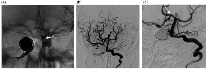
Results: We present five patients with symptomatic or ruptured PCA aneurysms with ICA occlusions. Age at presentation ranged from 21-58 and aneurysm size from 3-12 mm. All cases had angiographic evidence of posterior-anterior flow via the ipsilateral posterior communicating artery (PComA). The clinical presentation, diagnostic imaging and management strategies are further discussed. A literature review identified only two previous reported cases.

Conclusion: To our knowledge this is the first single centre series of posterior circulation aneurysms in patients with ICA occlusions that are considered to be 'flow-related.' The natural history of these rare lesions is unclear and the best management and surveillance strategy requires a patient-tailored approach by an experienced neurovascular team.

Keywords: Aneurysm; cerebral; flow-related; posterior circulation.


Conflict of interest statement

Declaration of conflicting interests: The author(s) declared no potential conflicts of interest with respect to the research, authorship, and/or publication of this article.

Figures

Figure 1.
Figure 1.
(a) Computed tomography angiogram (CTA) images demonstrated posteriorly projecting 3mm aneurysm (dashed black arrow) arising from a ‘hypertrophied’ left P1 posterior cerebral artery (PCA) segment (solid white arrow). (b) Severely stenosed distal cervical/laceral segments of the internal carotid arteries (ICA) bilaterally with (c) ‘beading’ appearances of the left vertebral consistent with fibromuscular dysplasia (FMD) (d) left vertebral and (e) external carotid artery (ECA) injections demonstrating filling of the internal carotid artery (ICA) via posterior-anterior flow via the posterior communicating artery (PComA) and via ECA collaterals (inferolateral trunk). (f) post-treatment appearances.
Figure 2.
Figure 2.
(a) Computed tomography angiogram (CTA) on initial presentation (5 years prior to the subarachnoid haemorrhage (SAH)) demonstrates normal appearances of the left posterior cerebral artery and (b) occlusion of the left internal carotid artery (ICA). (c) CTA after SAH revealed the culprit ipsilateral left P1/P2 posterior cerebral artery (PCA) 6mm aneurysm (white solid arrow). (d) Digital subtraction angiography (DSA) vertebral artery injection demonstrates the aneurysm and forward flow with filling of middle cerebral artery (MCA) branches visible (black solid arrow). (e) Post-treatment appearances.
Figure 3.
Figure 3.
(a) Three-dimensional-rotational angiography (3D-RA) images and (b) digital subtraction angiography (DSA) demonstrating the right P1/P2 junction posterior cerebral artery (PCA) aneurysm (dashed white arrow) ipsilateral to the internal cerebral artery (ICA) occlusion. A further 4mm aneurysm of the left superior cerebellar artery (SCA) origin was also noted (dashed black arrow). (b and c) Posterior-anterior flow into the middle cerebral artery (MCA) via the posterior communicating artery (PComA) was noted (solid white arrow). (c) Both aneurysms were treated with coil embolisation on the same occasion. (d) Two-year follow-up 3D-RA and e) DSA images revealed a 3mm neck recurrence (solid black arrow). (f) This was stent-coiled using a braided stent which was deployed in the PCA across the aneurysm neck resulting in complete occlusion.
Figure 4.
Figure 4.
(a) Computed tomography angiography (CTA) volume rendered image demonstrating the right P1 segment posterior cerebral artery (PCA) aneurysm. The distal P1 (dashed white arrow) and superior cerebellar artery (SCA) (solid white arrow) made the aneurysm not suitable for clipping or coil embolisation. (b) MRA minimal intensity projection image demonstrates the right superficial temporal artery to middle cerebral artery bypass appearance. Smaller calibre vertebrobasilar arteries are also noted following bilateral vertebral artery occlusion. (c) 25 year follow-up MRA image demonstrates stable appearances of the aneurysm.
Figure 5.
Figure 5.
(a) Left internal carotid artery (ICA) frontal run allows appreciation of one of the two left cavernous ICA aneurysms (solid white arrow). The coil mass on the right is related to prior treatment of a direct right carotico-cavernous fistula (CCF) and cavernous ICA aneurysm. (b) Left vertebral run demonstrating the right P1 (posterior cerebral artery) PCA aneurysm (solid black arrow) and forward flow through the posterior communicating artery (PComA) (dashed black arrow) which was treated with (c) stent assisted coiling (white dashed arrow).

Similar articles

Cited by

References

    1. Hassler O. Experimental carotid ligation followed by aneurysmal formation and other morphological changes in the circle of Willis. J Neurosurg 2009; 20: 1–7. DOI:10.3171/jns.1963.20.1.0001. - PubMed
    1. Gao L, Hoi Y, Swartz DD, et al.. Nascent aneurysm formation at the basilar terminus induced by hemodynamics. Stroke2008; 39: 2085–2090. DOI: 10.1161/STROKEAHA.107.509422. - PMC - PubMed
    1. Dyste GN, Beck DW. De novo aneurysm formation following carotid ligation: case report and review of the literature. Neurosurgery 1989; 24: 88–92. DOI: 10.1227/00006123-198901000-00014. - PubMed
    1. Fujiwara S, Fujii K, Fukui M.De novo aneurysm formation and aneurysm growth following therapeutic carotid occlusion for intracranial internal carotid artery (ICA) aneurysms. Acta Neurochir (Wien) 1993; 120: 20–25. - PubMed
    1. Wang YY, Rosenfeld JV, Lyon SM, et al.. Rapid development of a de novo intracranial aneurysm following carotid occlusion. J Clin Neurosci2008; 15: 324–330. DOI: 10.1016/j.jocn.2007.05.019. - PubMed

MeSH terms

LinkOut - more resources