Review
. 2021 Jun;46(6):502-517.
doi: 10.1016/j.tibs.2021.01.011. Epub 2021 Mar 3.

Direct Structural Insights into GABAA Receptor Pharmacology

Affiliations
Review

Direct Structural Insights into GABAA Receptor Pharmacology

Jeong Joo Kim et al. Trends Biochem Sci. 2021 Jun.

Abstract

GABAA receptors are pentameric ligand-gated ion channels that mediate most fast neuronal inhibition in the brain. In addition to their important physiological roles, they are noteworthy in their rich pharmacology; prominent drugs used for anxiety, insomnia, and general anesthesia act through positive modulation of GABAA receptors. Direct structural information for how these drugs work was absent until recently. Efforts in structural biology over the past few years have revealed how important drug classes and natural products interact with the GABAA receptor, providing a foundation for studies in dynamics and structure-guided drug design. Here, we review recent developments in GABAA receptor structural pharmacology, focusing on subunit assemblies of the receptor found at synapses.

Keywords: Cys-loop receptor structure; benzodiazepine; bicuculline; cryo-EM structure; general anesthetic; picrotoxin.


Conflict of interest statement

Declaration of Interests No interests are declared.

Figures
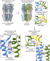
Figure 1:
Figure 1:. Historical progression of GABAA receptor structural biology.
Top left, side view of the X-ray structure of the β3 homomer (PDB code: 4COF). Top right, side view of the cryo-EM structure of the α1β2γ2 heteropentameric receptor and the binding sites for GABA (top) and flumazenil (bottom) (PDB code: 6D6U). Bottom right, the extracellular domain (ECD) and transmembrane domain (TMD) binding sites for diazepam in the α1β3γ2 receptor (PDB code: 6HUP). Bottom left, the binding site for propofol in the α1β2γ2 receptor (PDB code: 6X3T).
Figure 2:
Figure 2:. Neurotransmitter and benzodiazepine binding sites in the ECD.
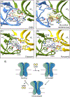
Panels A and B show the orthosteric neurotransmitter binding sites with the bound GABA (PDB code: 6X3Z) and bicuculline methbromide (PDB code: 6X3S), respectively. Panels C and D show the bound diazepam (PDB code: 6X3X) and flumazenil (PDB code: 6X3U), respectively, in the high affinity benzodiazepine site. Semitransparent surface is the experimental density map for the ligand. Panel E presents a cartoon schematic of the three principal conformational states of the receptor during its gating cycle.
Figure 3:
Figure 3:. Anesthetic and benzodiazepine binding sites in the TMD.
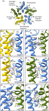
Panel A, synaptic perspective of the transmembrane domain (TMD) showing an overview of where different drugs were found to bind in the α1β2γ2 receptor structures. Panels B and C show the two distinct binding sites of TMD binding sites and ligand conformation for diazepam (PDB code: 6X3X). Panels D and E show the two binding sites identified for phenobarbital (PDB code: 6X3W). Panels F and G show representative binding sites for the IV anesthetics etomidate and propofol (PDB code: 6X3V and 6X3T), respectively.
Figure 4:
Figure 4:. TMD pore profiles for recent structures of the α1β2γ2 receptor.
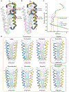
Panels A and B show the two binding sites for propofol (yellow) and etomidate (blue) in the context of the 4-helix bundle from individual β2 subunits. The emphasis is on how the conformation of the M2 helix differs among the GABA alone (grey), GABA + propofol, and GABA + etomidate structures. Panel C shows how pore diameter varies along the pseudo-5-fold channel axis: all three structures have a closed desensitization gate at the bottom of the pore, and variable constrictions at the 9’ activation gate. Panel D shows a pair of opposing M2 α-helices for each ligand complex; all structures include GABA bound except for bicuculline. Blue-green spheres and diameters illustrate shape of permeation pathway analyzed by HOLE (modified from Kim et al. 2020 [33]).
Figure I:
Figure I:. General architecture of a synaptic GABAA receptor.
Panel A, side view of the α1β2γ2 subunit assembly in complex with GABA (PDB code: 6X3Z). Panels B and C show the same structure from the perspective of the synapse of the extracellular domain (ECD) and transmembrane domain (TMD), respectively, highlighting binding sites for different small molecules.

References

    1. Tasneem A, et al., Identification of the prokaryotic ligand-gated ion channels and their implications for the mechanisms and origins of animal Cys-loop ion channels. Genome Biol, 2005. 6(1): p. R4. - PMC - PubMed
    1. Jaiteh M, Taly A, and Henin J, Evolution of Pentameric Ligand-Gated Ion Channels: Pro-Loop Receptors. PLoS One, 2016. 11(3): p. e0151934. - PMC - PubMed
    1. Nemecz A, et al., Emerging Molecular Mechanisms of Signal Transduction in Pentameric Ligand-Gated Ion Channels. Neuron, 2016. 90(3): p. 452–70. - PubMed
    1. Sieghart W and Savic MM, International Union of Basic and Clinical Pharmacology. CVI: GABAA Receptor Subtype- and Function-selective Ligands: Key Issues in Translation to Humans. Pharmacol Rev, 2018. 70(4): p. 836–878. - PubMed
    1. Sigel E and Ernst M, The Benzodiazepine Binding Sites of GABAA Receptors. Trends Pharmacol Sci, 2018. 39(7): p. 659–671. - PubMed

Publication types

MeSH terms

Substances

LinkOut - more resources