Review
. 2021 Sep;46(9):744-757.
doi: 10.1016/j.tibs.2021.01.014. Epub 2021 Mar 3.

Rapid Detection and Signaling of DNA Damage by PARP-1

Affiliations
Review

Rapid Detection and Signaling of DNA Damage by PARP-1

Nootan Pandey et al. Trends Biochem Sci. 2021 Sep.

Abstract

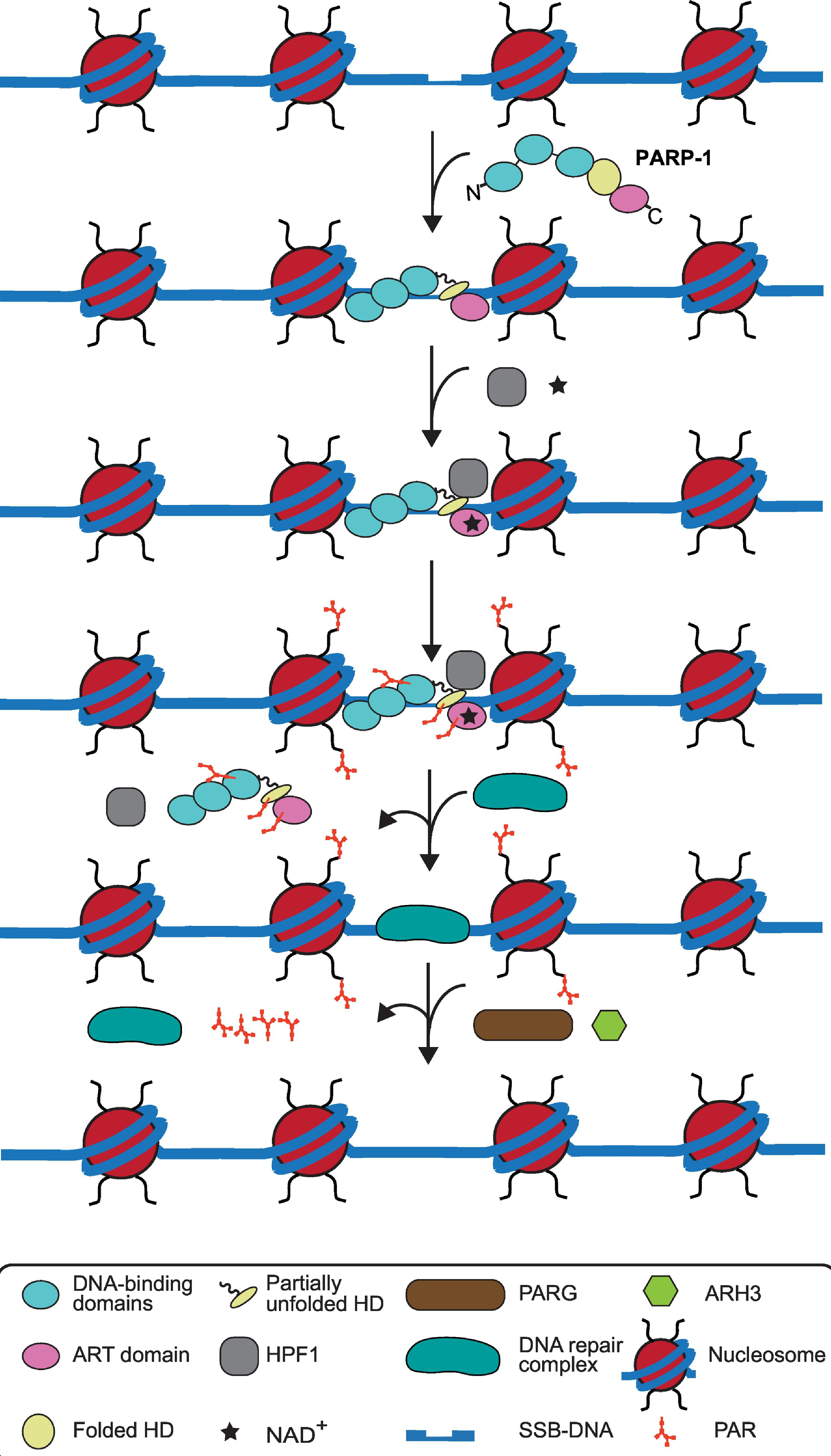
Poly(ADP-ribosyl) polymerase-1 (PARP-1) is an abundant ADP-ribosyl transferase that regulates various biological processes. PARP-1 is widely recognized as a first-line responder molecule in DNA damage response (DDR). Here, we review the full cycle of detecting DNA damage by PARP-1, PARP-1 activation upon DNA binding, and PARP-1 release from a DNA break. We also discuss the allosteric consequence upon binding of PARP inhibitors (PARPi) and the opportunity to tune its release from a DNA break. It is now possible to harness this new understanding to design novel PARPi for treating diseases where cell toxicity caused by PARP-1 'trapping' on DNA is either the desired consequence or entirely counterproductive.

Keywords: DNA damage response; PARPi; PARylation; poly(ADP-ribose) glycohydrolase.


Conflict of interest statement

Declaration of Interests B.E.B. is a founder of Hysplex LLC, with interests in PARPi development.

Figures

Figure 1
Figure 1. Structure of PARP-1 on a DNA break and relationship to other DNA damage PARPs.
(A) Domains of human PARP-1. The N-terminal domain includes zinc-fingers (F1, F2, and F3), followed by automodification domain (AD) which has automodification sites. The DNA binding (WGR) domain and catalytic (CAT) domain resides at C-terminal domain. The helical domain (HD) and ADP-ribosyl transferase (ART) domain comprise the CAT domain. (B) Combined model of the crystal structure of F1, F3, and WGR-CAT with DNA complex (PDB:4DQY) and NMR structure of F1-F2 with an SSB [25]. (C) Domain organization of the three DNA damage PARPs. The N-terminal region (NTR) of PARP-1 has three zinc-fingers (F1, F2, and F3) and BRCT domain whereas PARP-2 and PARP-3 has shorter NTR and lack zinc-fingers. All three DNA damage PARPs share WGR, HD, and ART domain. (D) Similarities and differences in the biochemical activities of the three DNA damage PARPs. BER: base excision repair, SSBR: single strand break repair, DBSR: double strand break repair, HR: homologous recombination and NHEJ: non-homologous end joining.
Figure 2
Figure 2. Simplified cycle of PARP-1 on and off of a DNA break.
In the absence of DNA damage, PARP-1 domains are unconnected, resembling a beads-on-a-string arrangement. The helical domain (HD) is an autoinhibitory domain and regulates PARP-1 catalytic activity via blocking the NAD+ binding to the nicotinamide site (N) of the ADP-ribosyl transferase (ART) domain. PARP-1 detects DNA damage through its DNA binding domains (zinc fingers F1, F2, and F3) and collapses into a compact conformation around the broken DNA lesion. The resulting interdomain contacts destabilize the HD (double headed arrow), permitting NAD+ ready access to N. NAD+ binding reciprocally strengthens the PARP-1 interaction with broken DNA through reverse allosteric communication from catalytic domain to DNA binding domains (single-headed arrow). PARP-1 automodification, predominantly at the BRCT automodification domain (AD) permits PARP-1 release from DNA.
Figure 3
Figure 3. The cycle of PARP-1 in DDR signaling in the context of chromatin, HPF1, downstream repair factors, and PAR hydrolyzing enzymes.
PARP-1 detects and binds to damaged DNA with allosteric changes in helical domain (HD) domain. HPF1, an accessory factor, binding to the catalytic domain and NAD+ access results in catalytic activation of PARP-1. PARP-1 and HPF1 are thought to form a composite active site for PARylation of PARP-1 and histones present in nucleosomes in the vicinity of DNA break. PARP-1 automodification leads to its release from DNA. The removal of PARP-1 and PAR residues on histones occurs simultaneously with the recruitment of DNA repair complex, consisting of XRCC1, APTX, PNKP, DNA Polymeraseβ, and Ligase 3α. Lastly, after the DNA break is repaired, then the repair complex is dissociated from DNA and PARG and ARH3 remove local PAR chains.

References

    1. Gibson BA and Kraus WL (2012) New insights into the molecular and cellular functions of poly(ADP-ribose) and PARPs. Nat. Rev. Mol. Cell. Biol 13, 411–424 - PubMed
    1. Amé JC et al. (2004) The PARP superfamily. Bioessays 26, 882–893 - PubMed
    1. Chambon JD et al. (1963) Nicotinamide mononucleotide activation of new DNA-dependent polyadenylic acid synthesizing nuclear enzyme. Biochem. Biophys. Res. Commun 11, 39–43 - PubMed
    1. Amé JC et al. (1999) PARP-2, A novel mammalian DNA damage-dependent poly(ADP-ribose) polymerase. J. Biol. Chem 274, 17860–17868 - PubMed
    1. Rulten SL et al. (2011) PARP-3 and APLF function together to accelerate nonhomologous end-joining. Mol. Cell 41, 33–45 - PubMed

Publication types

MeSH terms

Substances

LinkOut - more resources