Case Reports
. 2021 Mar 5;14(3):e235676.
doi: 10.1136/bcr-2020-235676.

Management of severe traumatic intrusion in the permanent dentition

Affiliations
Case Reports

Management of severe traumatic intrusion in the permanent dentition

Alex Rovira-Wilde et al. BMJ Case Rep. .

Abstract

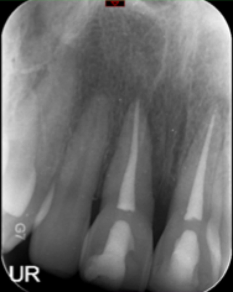
Traumatic intrusion is considered one of the most severe luxation injuries to the permanent dentition. There are limited studies based on minimal evidence supporting suggested management protocols, owing to the rare occurrence of intrusion. The following case report details the multidisciplinary management and 18-month follow-up, in line with current UK guidelines, of a 23-year old adult male who sustained severe intrusion injuries to both permanent maxillary central incisor teeth. Timely, accurate diagnosis and subsequent appropriate management correlates with improved outcomes for traumatic injuries and it is therefore imperative those involved with the acute and long-term management of dentoalveolar trauma are aware of current guidelines.

Keywords: accidents; dentistry and oral medicine; injuries.


Conflict of interest statement

Competing interests: None declared.

Figures

Figure 1
Figure 1
Initial presentation 36 hours following injury. Incisal edge of maxillary central incisors partially visible.
Figure 2
Figure 2
Standard maxillary occlusal radiograph identifying apical displacement of the maxillary central incisors (36 hours following injury).
Figure 3
Figure 3
Periapical radiographs suggesting apical displacement of the maxillary central incisors (36 hours following injury).
Figure 4
Figure 4
An orthopantomogram confirming severe apical displacement of the maxillary central incisors and ruled out facial fractures taken within the accident and emergency department following trauma (radiolucency suggestive of caries to be managed by the general dental practitioner).
Figure 5
Figure 5
Provision of titanium trauma splint following surgical repositioning of maxillary central incisors (36 hours following injury).
Figure 6
Figure 6
Periapical radiographs showing maxillary central incisors immediately following endodontic treatment (7 weeks following injury).
Figure 7
Figure 7
Eighteen-month postoperative clinical views, evidence of marginal bone loss around maxillary central incisors present interproximally.
Figure 8
Figure 8
Periapical radiographs showing maxillary central incisors at 6-month (left) and 18-month (right) review following endodontic treatment.

References

    1. Andreasen JO, Bakland LK, Matras RC, et al. . Traumatic intrusion of permanent teeth. Part 1. An epidemiological study of 216 intruded permanent teeth. Dent Traumatol 2006;22:83–9. 10.1111/j.1600-9657.2006.00421.x - DOI - PubMed
    1. Petersson EE, Andersson L, Sörensen S. Traumatic oral vs non-oral injuries. Swed Dent J 1997;21:55–68. - PubMed
    1. Wigen TI, Agnalt R, Jacobsen I. Intrusive luxation of permanent incisors in Norwegians aged 6-17 years: a retrospective study of treatment and outcome. Dent Traumatol 2008;24:612–8. 10.1111/j.1600-9657.2008.00686.x - DOI - PubMed
    1. Diangelis AJ, Andreasen JO, Ebeleseder KA, et al. . International association of dental Traumatology guidelines for the management of traumatic dental injuries: 1. fractures and luxations of permanent teeth. Dent Traumatol 2012;28:2–12. 10.1111/j.1600-9657.2011.01103.x - DOI - PubMed
    1. Albadri S, Zaitoun H, Kinirons MJ, et al. . Uk national clinical guidelines in paediatric dentistry: treatment of traumatically intruded permanent incisor teeth in children. Int J Paediatr Dent 2010;20 Suppl 1:1–2. 10.1111/j.1365-263X.2010.01085.x - DOI - PubMed

Publication types