. 2021 Mar 5;7(10):eabf2403.
doi: 10.1126/sciadv.abf2403. Print 2021 Mar.

The mycoplasma surface proteins MIB and MIP promote the dissociation of the antibody-antigen interaction

Affiliations

The mycoplasma surface proteins MIB and MIP promote the dissociation of the antibody-antigen interaction

Pierre Nottelet et al. Sci Adv. .

Abstract

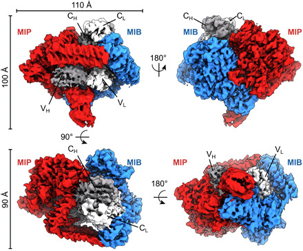
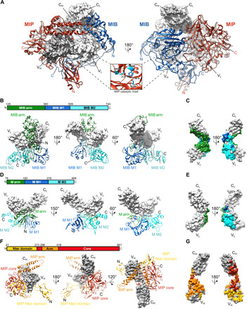
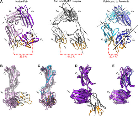
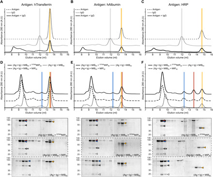
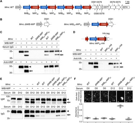
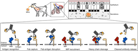
Mycoplasma immunoglobulin binding (MIB) and mycoplasma immunoglobulin protease (MIP) are surface proteins found in the majority of mycoplasma species, acting sequentially to capture antibodies and cleave off their VH domains. Cryo-electron microscopy structures show how MIB and MIP bind to a Fab fragment in a "hug of death" mechanism. As a result, the orientation of the VL and VH domains is twisted out of alignment, disrupting the antigen binding site. We also show that MIB-MIP has the ability to promote the dissociation of the antibody-antigen complex. This system is functional in cells and protects mycoplasmas from antibody-mediated agglutination. These results highlight the key role of the MIB-MIP system in immunity evasion by mycoplasmas through an unprecedented mechanism, and open exciting perspectives to use these proteins as potential tools in the antibody field.


Figures

Fig. 1
Fig. 1. Cryo-EM density map of the tripartite complex between MIB83, S759AMIP82, and a goat IgG Fab.
The map is segmented with MIB83 colored in blue, S759AMIP82 in red, the Fab heavy chain in dark gray, and the Fab light chain in light gray. Alternative orientations of the map are shown, and the axis and angles of rotation between these orientations are indicated, as well as the overall dimensions of the complex.
Fig. 2
Fig. 2. Structures of MIB83 and S759AMIP82 and their interactions with the Fab.
(A) Structure of the complex between MIB83, S759AMIP82, and a goat IgG Fab fragment (gray surfaces). Inset: zoomed-in view of the catalytic triad in the S759AMIP82 core domain. (B) Interaction of MIB83 (ribbon) with the Fab light chain (surface). MIB83 structure is split into three domains: MIB arm (green), MIB M1 (blue), and MIB M2 (cyan). (C) Footprint of MIB83 on the Fab light chain in surface representation. (D) Structure of Protein M [ribbon; Protein Data Bank (PDB): 4NZR] in complex with the light chain of a Fab (gray surface). Protein M structure is split into three domains: M arm (green), M M1 (blue), and M M2 domains (cyan). (E) Footprint of Protein M on the light chain of the Fab in surface representation. (F) Interaction of S759AMIP82 (ribbon) with the Fab heavy chain (gray surface). S759AMIP82 is split into three domains: MIP N-terminal domain (yellow), MIP core (red), and MIP arm domains (orange). (G) Footprint of S759AMIP82 on the Fab heavy chain in surface representation.
Fig. 3
Fig. 3. The structure of the antigen binding site of the Fab bound to MIB83 and S759AMIP82 is disrupted.
(A) Comparison of isolated and bound Fabs structures. Left: Isolated Fab (PDB: 6TCM). Light chain: pink; heavy chain: purple. Middle: Goat IgG Fab in complex with MIB83 and S759AMIP82. Light chain: light gray; heavy chain: dark gray. Right: Fab in complex with Protein M (PDB: 4NZR). Light chain: cyan; heavy chain: blue. The main axis of each domain was determined in Chimera and is indicated as a black dashed line. The calculated angles between these axes are displayed, as well as the distance between the CDR1 loops of the VL and VH domains. The complementarity-determining regions (CDRs) of the VH and VL domains are colored orange and yellow, respectively. (B to D) Superimposition of the structures of various isolated Fabs (PDB: 6TCM, 4POZ, 4HBC, 4POZ, and 5ILT) with the Fab bound to MIB83-S759AMIP82 and to Protein M. (B) Superimposition of the light chains of isolated Fabs and Fab bound to MIB-MIP. (C) Superimposition of the light chains of isolated Fabs and Fab bound to Protein M. (D) Superimposition of the heavy chains of isolated Fabs and Fab bound to MIB-MIP. (E) Superimposition of the heavy chains of isolated Fabs and Fab bound to Protein M.
Fig. 4
Fig. 4. MIB and MIP promote the dissociation of the antigen-antibody interaction.
(A to C) Size exclusion chromatography analysis of the interaction between a specific antibody (IgG) and its corresponding antigen [(A) hTransferrin; (B) hAlbumin; (C) HRP]. The IgG and the antigen were incubated for 30 min before injection in the column. Colored vertical bars indicate the specific elution volumes of the antigen (gold) and the IgG (gray). (D to F) Interaction between the IgG and its corresponding antigen [(D) hTransferrin; (E) hAlbumin; (F) HRP] in the presence of MIB-MIP or MIB alone. Top: Size exclusion chromatography analysis of the different samples. Colored vertical bars indicate the specific elution volumes of MIB83 (blue), MIP82 and S759AMIP82 (red), and the antigens (gold). Bottom: SDS-PAGE analysis of the different fractions collected during the size exclusion chromatography. Arrowheads indicate the position of each protein: MIB83 (blue), MIP82 or S759AMIP82 (red), IgG heavy chain (dark gray), IgG light chain (light gray), and the antigens (gold). The star indicates the cleavage of the IgG heavy chain. For the antigen HRP, two bands can be seen for the cleaved IgG heavy chain due to the polyclonal nature of the anti-HRP antibodies used here. A.U., arbitrary units.
Fig. 5
Fig. 5. MIB and MIP are active in vivo and protect the cells from immunoglobulins targeting surface antigens.
(A) Schematic representation of the Mmc genomic locus encoding MIBs (blue) and MIPs (red) homologs. (B) Genomic loci encoding MIBs and MIPs in the Mmc mutant strains ΔMIB-MIP and MIB83-MIP82. (C) Western blot analysis of immunoglobulin cleavage by the wild-type and mutant Mmc cells. The assays were performed with nonspecific polyclonal IgG from normal goat serum or anti-HRP polyclonal IgG affinity-purified from immunized goat. Arrowheads indicate the location of the intact or cleaved (star) immunoglobulin heavy chain. (D) Top: Schematic representation of the MIB-MIP locus in the mutant Mmc MIP82-HA. Bottom: Western blot analysis of anti-HA goat IgG cleavage by wild-type and mutant Mmc cells. (E) Analysis of immunoglobulin cleavage performed using sera collected from goats infected by Mmc (“D0”: before infection; “D12”: 12 days after infection). Each sample was split into two and analyzed by Western blot against goat IgM or goat IgM. (F) Agglutination of wild-type and mutant Mmc cells by the sera of infected goats. Cells were cultivated in the presence of 2% goat serum and agglutination was observed by dark-field microscopy or quantified in microplates by measuring the absorbance at 310 nm.
Fig. 6
Fig. 6. Model of the MIB-MIP mechanism.
Mycoplasma enters the airways of the host through airborne droplets or nose-to-nose contacts and will migrate to the lower portions of the lungs to establish its infection. The lung epithelium is lined with secretions, including the pulmonary surfactant, that are rich in secretory IgA and also contain small amounts of IgG and IgM. MIB (blue) is located at the mycoplasma cell surface and is able to target antibodies that are specifically bound to a surface antigen (beige). MIB binds to the light chain of the antibody and displaces the antibody from its cognate antigen. The antigen binding site conformation of the antibody is disrupted as the VH domain of the immunoglobulin is shifted away from its initial position. MIP (red) is subsequently recruited and activated, resulting in the cleavage of the VH domain. The broken immunoglobulin is then released from the MIB-MIP complex.

Similar articles

Cited by

References

    1. Kuijl C., Neefjes J., New insight into the everlasting host-pathogen arms race. Nat. Immunol. 10, 808–809 (2009). - PubMed
    1. Chaplin D. D., Overview of the immune response. J. Allergy Clin. Immunol. 125, S3–S23 (2010). - PMC - PubMed
    1. Schroeder H. W. J. Jr., Cavacini L., Structure and function of immunoglobulins. J. Allergy Clin. Immunol. 125, S41–S52 (2010). - PMC - PubMed
    1. Finlay B. B., McFadden G., Anti-immunology: Evasion of the host immune system by bacterial and viral pathogens. Cell 124, 767–782 (2006). - PubMed
    1. Sidorin E. V., Solov’eva T. F., IgG-binding proteins of bacteria. Biochemistry (Mosc.) 76, 295–308 (2011). - PubMed

Publication types