. 2021 Mar 5;12(1):1483.
doi: 10.1038/s41467-021-21737-9.

Cytotoxic CD8+ T cells promote granzyme B-dependent adverse post-ischemic cardiac remodeling

Affiliations

Cytotoxic CD8+ T cells promote granzyme B-dependent adverse post-ischemic cardiac remodeling

Icia Santos-Zas et al. Nat Commun. .

Abstract

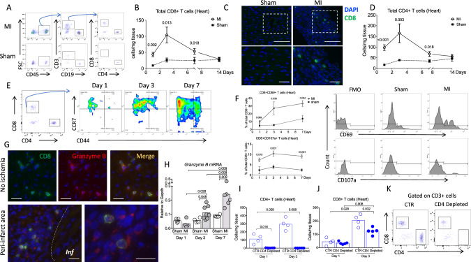
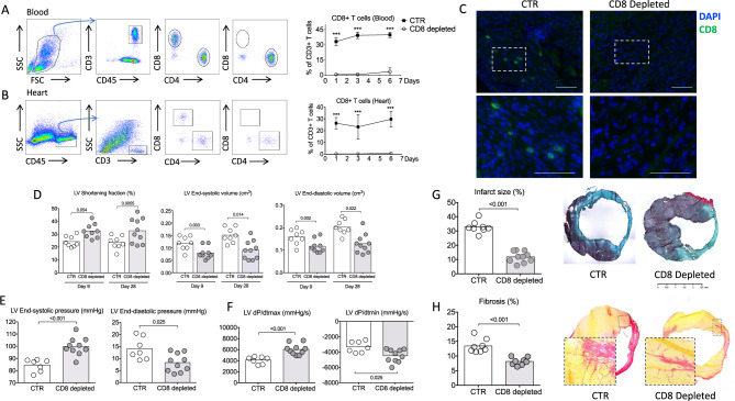
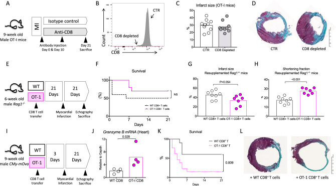
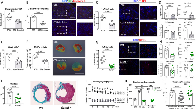
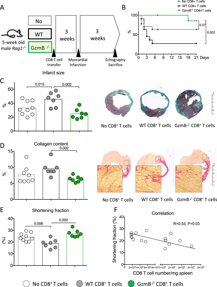
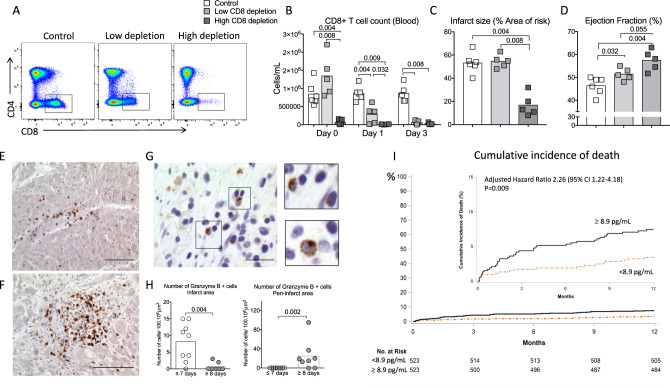
Acute myocardial infarction is a common condition responsible for heart failure and sudden death. Here, we show that following acute myocardial infarction in mice, CD8+ T lymphocytes are recruited and activated in the ischemic heart tissue and release Granzyme B, leading to cardiomyocyte apoptosis, adverse ventricular remodeling and deterioration of myocardial function. Depletion of CD8+ T lymphocytes decreases apoptosis within the ischemic myocardium, hampers inflammatory response, limits myocardial injury and improves heart function. These effects are recapitulated in mice with Granzyme B-deficient CD8+ T cells. The protective effect of CD8 depletion on heart function is confirmed by using a model of ischemia/reperfusion in pigs. Finally, we reveal that elevated circulating levels of GRANZYME B in patients with acute myocardial infarction predict increased risk of death at 1-year follow-up. Our work unravels a deleterious role of CD8+ T lymphocytes following acute ischemia, and suggests potential therapeutic strategies targeting pathogenic CD8+ T lymphocytes in the setting of acute myocardial infarction.


Conflict of interest statement

H. Ait-Oufella applied a patent on the protective effect of CD8 depletion and Granzyme B blockade after acute myocardial infarction. The other authors declare no competing interests.

Figures

Fig. 1
Fig. 1. Cytotoxic CD8+ T lymphocytes are activated and recruited to the ischemic tissue after myocardial infarction.
A Representative examples of CD4+ and CD8+ T cell staining in the heart of C57BL/6J mice following coronary ligation (MI) or sham surgery. B Kinetic of CD8+ T cell infiltration in the myocardium, performed at days 0, 1, 3, 7, and 14 after surgery in MI (white circle, n = 9/12/12/5, respectively, at day 1/3/7/14) and sham-operated mice (black box, n = 6/time point); data are presented as mean ± SEM. C Immunostaining in the ischemic myocardium at day 3 after MI or sham showing CD8+ T infiltration (green) in the ischemic heart tissue, scale bar 40 μm. D Kinetic of CD4+ T cell infiltration in the myocardium, performed at days 0, 1, 3, 7, and 14 after surgery in MI (white circle, n = 10/9/12/5, respectively, at day 1/3/7/14) and sham-operated mice (black box, n = 6/time point); data are presented as mean ± SEM. E Flow cytometry characterization of CD8+ T cell subsets in the ischemic heart at days 1, 3, and 7 after MI including naïve (CD3+CD8+CCR7HighCD44), effector memory (CD3+CD8+CCR7LowCD44+), and central memory (CD3+CD8+CCR7HighCD44+) subsets. F Representative examples and quantitative analysis of CD8+ T cells expressing CD69 or CD107a within the ischemic heart tissue in MI (white circle, n = 6/9/12, respectively, at day 1/3/7) and sham-operated mice (black box, n = 6/time point); data are presented as mean ± SEM. G Immunostaining in the ischemic myocardium at day 3 after MI showing CD8+ T cells (green), and Granzyme B (Red) and merged area (Yellow), Inf for infarct area; scale bar 40 μm. H mRNA levels of Granzyme B within the injured myocardium on days 1, 3, and 7 after coronary ligation (gray, n = 5/4/5 at day 1/3/7) or sham (white, n = 4/8/6 at day 1/3/7). I C57Bl6 WT mice received intraperitoneal injection of isotype control (CTR, blue borderline, n = 4/time point) or anti-CD4 depleting monoclonal antibody (blue filled, n = 5/time point) (150 μg/mice) 1 day before coronary occlusion. CD4+ T cell depletion was confirmed in the heart by flow cytometry at day 1 and day 3 following MI. J, K Quantification and representative example of CD8+ T cell count in the heart of control (n = 4/time point) or CD4-depleted (n = 5/time point) mice at day 1 and day 3 after MI. P values were calculated using two-tailed Mann-Whitney test (B, D, F, H, I, J). Inf, infarct; MI, myocardial infarction; FMO, fluorescent minus one.
Fig. 2
Fig. 2. CD8 T cell depletion improves heart function and reduces infarct size.
A Representative examples (left) and quantitative analysis (right) of CD8+ T cell staining in the blood of C57BL/6J mice treated with isotype control (CTR, black box) or with the CD8 mAb (CD8 depleted, white circle) (n = 6 mice per group/time point); data are presented as mean ± SEM, ***P < 0.001. B Representative examples (left) and quantitative analysis (right) of CD8+ T cell staining in the heart of C57BL/6J mice treated with isotype control (CTR) or with the CD8 mAb (CD8 depleted) (n = 6 mice per group/time point); data are presented as mean ± SEM, ***P < 0.001. C Representative examples of CD8+ T cell staining in the peri-infarct area of C57BL/6J mice treated with isotype control (CTR) or with the CD8 mAb (CD8 depleted) at day 3, scale bar 40 μm. D Echocardiography analysis after anti-CD8 therapy. We measured LV shortening fraction (SF), LV volume at end systole and end diastole *(CTR white, n = 8 and CD8 depleted gray n = 9–10). E Systolic and diastolic pressure measured in the LV, and F related function parameters using intracardiac probe at day 28 (CTR n = 7 and CD8 depleted n = 9–10). G Representative photomicrographs and quantitative analysis of infarct size evaluation evaluated by Masson trichrome staining, in the 2 groups of mice (CTR n = 8 and CD8 depleted n = 10). H Representative photomicrographs and quantitative analysis of myocardial fibrosis evaluated by Sirius Red staining, in the 2 groups of mice (CTR n = 8 and CD8 depleted n = 9). LV, left ventricle. P values were calculated using two-tailed Mann-Whitney test (A, B, D, E, F, G, H).
Fig. 3
Fig. 3. CD8+ T cells pathogenic activity requires antigen-specific stimulation.
A Male 9-week-old OT-I mice were treated with isotype control (CTR, white) or the CD8 mAb (CD8 depleted, gray). B CD8 depletion (red) was confirmed in the spleen at day 21 using flow cytometry. C, D Representative photomicrographs and quantitative analysis of infarct size evaluation using Masson trichrome staining, in the 2 groups of OT-I mice (CTR n = 10 and CD8 depleted n = 9). E Rag1−/− mice injected with CD8-depleted splenocytes re-supplemented with WT (white) or OT-I (pink) CD8+ T cells, 3 weeks before MI. F Survival rate following MI (from 2 experiments, WT n = 16 and OT-I n = 13). G Quantitative analysis of infarct size evaluation assessed by Masson trichrome staining in the 2 groups of mice (WT n = 8 and OT-I n = 7). H Echocardiography analysis 21 days after MI and assessment of LV shortening fraction (SF) in the 2 groups of re-supplemented mice (WT n = 8 and OT-I n = 7). I CMy-mOva mice were injected with WT (white) or OT-I (pink) CD8+ T cells, 3 days before MI. J Granzyme B mRNA expression in the ischemic heart at day 2 after MI in CMy-mOva mice injected with WT or OT-I CD8+ T cells (n = 4/group). K Survival rate following MI (pooled 2 experiments, WT n = 10 and OT-I n = 12). L Representative examples of infarct size after Masson trichrome staining. P values were calculated using two-tailed Mann-Whitney test (C, G, H, J). Difference in survival was evaluated using log-rank test (F, K).
Fig. 4
Fig. 4. CD8+ T lymphocyte depletion or Granzyme B global deficiency reduces cardiomyocyte apoptosis and pro-inflammatory responses within the ischemic heart tissue.
A Representative histograms of mRNA levels of Granzyme B within the injured myocardium on day 3 after MI in CTR (white) and CD8-depleted (gray) mice (n = 5/group). B Representative examples (right) and quantitative analysis (left) of Granzyme B staining in the ischemic heart of C57BL/6J mice with or without CD8 depletion (n = 5/group); scale bars 50 and 25 μm. C Representative examples (right) and quantitative analysis (left) of TUNEL+ cells (Red) in the peri-infarct area of C57BL/6J mice (n = 5/group); scale bars 50 and 25 μm. D Representative histograms of mRNA levels of Il-1β, I-6, Tnf-α, and Il-10 within the injured myocardium on day 7 after MI (n = 5/group). E Representative histograms of mRNA levels of Mmp9 within the injured myocardium on day 7 after MI (n = 5/group). F Quantification (left) and representative photomicrographs (right) of matrix metalloproteinase (MMP)-sense 680 activity in the ischemic heart measured by ex vivo reflectance epifluorescence imaging at day 7 (CTR n = 5 and CD8 depleted n = 6), scale bar 2.5 mm. G Acute MI was induced on C57bl6 wild-type (WT, white) mice or Granzyme B deficient (GzmB−/−, green) mice. Representative examples (right) and quantitative analysis (left) of TUNEL+ cells in the peri-infarct area of WT C57BL/6J or GzmB−/− mice at day 3 after MI (WT n = 8 and GzmB−/− n = 9); scale bars 50 and 25 μm. H Il-1β, Il-6, Tnf-α, and Il-10 mRNA levels measured by qPCR in infarcted heart at day 3 after MI (n = 9/group). I Representative photomicrographs (left) and quantitative analysis (right) of infarct size evaluation using Masson trichrome staining, in the 2 groups of mice (n = 7/group); scale bar 2.5 mm. J Purified non-activated or activated WT CD8+ T cells were co-cultured with cardiomyocytes at different ratios (Cardiomyocyte/CD8+ T cells) for 24 h before their removal. Apoptotic cardiomyocytes labeled with an active caspase-3 fluorescent dye was monitored for 24 h (n = 4–5/conditions), Data are presented as mean ± SEM, #P < 0.001 for activated CD8+ T cells 1/5 versus non-activated CD8+ T cells or activated CD8+ T cells 1/1 or activated CD8+ T cells 1/3; #P < 0.001 for activated CD8+ T cells 1/10 versus non-activated CD8+ T cells or activated CD8+ T cells 1/1 or activated CD8+ T cells 1/3. K WT or GzmB−/− CD8+ T cells were co-cultured with cardiomyocytes for 24 h at 1/5 and 1/10 ratio and cardiomyocyte apoptosis using an active caspase-3 fluorescent dye was quantified. Cardiomyocyte and non-activated CD8+ T cells co-culture was named CTR condition (n = 4/condition) ***P < 0.001. L Isolated cardiomyocytes were co-cultured overnight with CD8+ T cells isolated from WT or GzmB−/− mice and cardiomyocyte sarcomere shortening was measured (non-activated n = 14, activated n = 17 and GzmB−/− n = 10) at a ratio 1/3. P values were calculated using two-tailed Mann-Whitney test (A, B, C, D, E, F, G, H, I, J) or Kruskal-Wallis test (K, L). TUNEL, terminal deoxynucleotidyl transferase dUTP Nick End Labeling; MMP, matrix metalloprotease.
Fig. 5
Fig. 5. CD8+ T lymphocytes trigger adverse ventricular remodeling and alter heart function through the production of Granzyme B.
A Rag1−/− mice injected with either CD8-depleted splenocytes (White) or CD8 cell-depleted splenocytes re-supplemented with WT (gray) or GzmB−/− CD8+ T cells (green), 3 weeks before MI. B Survival curves following MI (from 3 experiments, No CD8 n = 15, WT CD8 n = 18, and GzmB−/− CD8 n = 8). C Representative photomicrographs and quantitative analysis of infarct size. D Collagen content in the peri-infarct area in the 3 groups of mice, scale bar 100 μm. Results are pooled from three independent experiments including surviving mice (no CD8 n = 9, WT CD8 n = 7, and GzmB−/− CD8 n = 7). E Echocardiography analysis after 21 days of MI and assessment of LV shortening fraction (SF) in the 3 groups of mice (no CD8 n = 9, WT CD8 n = 7, and GzmB−/− CD8 n = 7). F Correlation between CD8+ T cell number in the spleen at day 21 and LV shortening fraction. Data from CD8-depleted splenocytes or CD8 cell-depleted splenocytes re-supplemented with WT CD8+ T cells have been included (n = 16). P values were calculated using two-tailed Kruskal-Wallis test (C, D, E). Difference in survival was evaluated using log-rank test (B) and correlation was studied using Spearman’s test (F).
Fig. 6
Fig. 6. Pathogenic role of CD8+ T cells in a model of myocardial ischemia/reperfusion in pig and relevance to the human disease.
A Flow cytometry analysis of blood CD4+ T and CD8+ T subsets at day 1 after MI in control (PBS, CTR, n = 6), low (light gray, n = 5), and high (dark gray, n = 5) CD8-depleted groups. B Quantification of CD8+ T cell count in the blood at baseline, day 0, day 1, and day 3 after MI. C Representative picture and quantification of infarct size at day 14 in control, low, and high CD8-depleted groups. D Quantitative evaluation of left ventricle ejection fraction (Simpson) of CTR or CD8-depleted pigs. E, F Detection of CD8+ T cells (brown) in human heart biopsy of MI patients, using immunohistochemistry at day 3 (E, upper) and day 8 (F, lower) after MI, scale bar 50 μm. G Detection of GRANZYME B+ cells (brown) in human heart biopsy of MI patients, using immunohistochemistry, scale bar 20 μm. H Quantification of GRANZYME B+ cells (brown) in human heart biopsy of MI patients, using immunohistochemistry at different time points (n = 9 day ≤ 7, n = 8 day ≥ 8). I Survival according to baseline circulating GRANZYME B level (< or > median value) in patients with acute MI (n = 1046). High level of GRANZYME B at the admission for acute MI were independently predictive of death after 1 year of follow-up after multiple adjustments (see Methods and Supplementary Table 3). HR = hazard ratio. P values were calculated using two-tailed Mann-Whitney test (H) or Kruskal-Wallis test (BD).

Similar articles

Cited by

References

    1. Frangogiannis NG. The inflammatory response in myocardial injury, repair, and remodelling. Nat. Rev. Cardiol. 2014;11:255–265. doi: 10.1038/nrcardio.2014.28. - DOI - PMC - PubMed
    1. Rosamond WD, et al. Twenty-two-year trends in incidence of myocardial infarction, coronary heart disease mortality, and case fatality in 4 US communities, 1987–2008. Circulation. 2012;125:1848–1857. doi: 10.1161/CIRCULATIONAHA.111.047480. - DOI - PMC - PubMed
    1. Puymirat E, et al. Association of changes in clinical characteristics and management with improvement in survival among patients with ST-elevation myocardial infarction. JAMA. 2012;308:998–1006. doi: 10.1001/2012.jama.11348. - DOI - PubMed
    1. Lavoie L, Khoury H, Welner S, Briere JB. Burden and prevention of adverse cardiac events in patients with concomitant chronic heart failure and coronary artery disease: a literature review. Cardiovasc. Ther. 2016;34:152–160. doi: 10.1111/1755-5922.12180. - DOI - PMC - PubMed
    1. Nahrendorf M. Myeloid cell contributions to cardiovascular health and disease. Nat. Med. 2018;24:711–720. doi: 10.1038/s41591-018-0064-0. - DOI - PMC - PubMed

Publication types

MeSH terms

LinkOut - more resources