. 2021 Apr;40(14):2496-2508.
doi: 10.1038/s41388-021-01710-y. Epub 2021 Mar 5.

Molecular disruption of DNA polymerase β for platinum sensitisation and synthetic lethality in epithelial ovarian cancers

Affiliations

Molecular disruption of DNA polymerase β for platinum sensitisation and synthetic lethality in epithelial ovarian cancers

Reem Ali et al. Oncogene. 2021 Apr.

Abstract

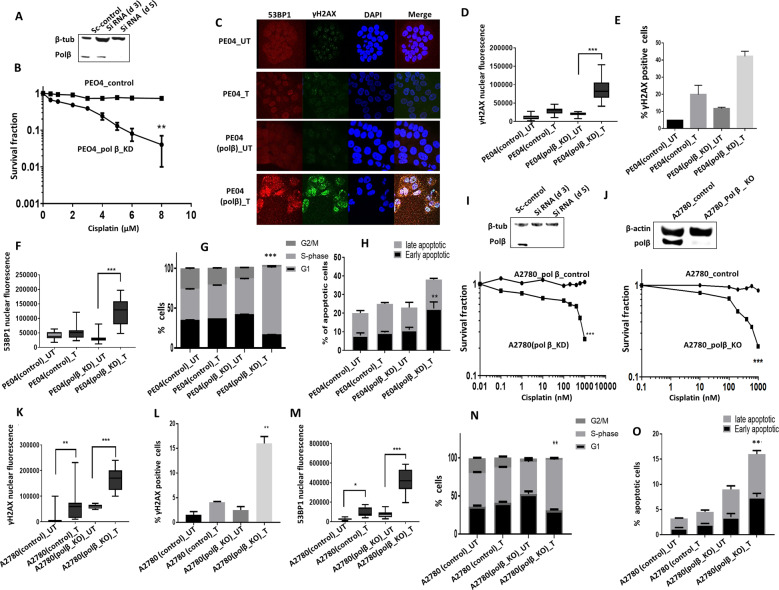
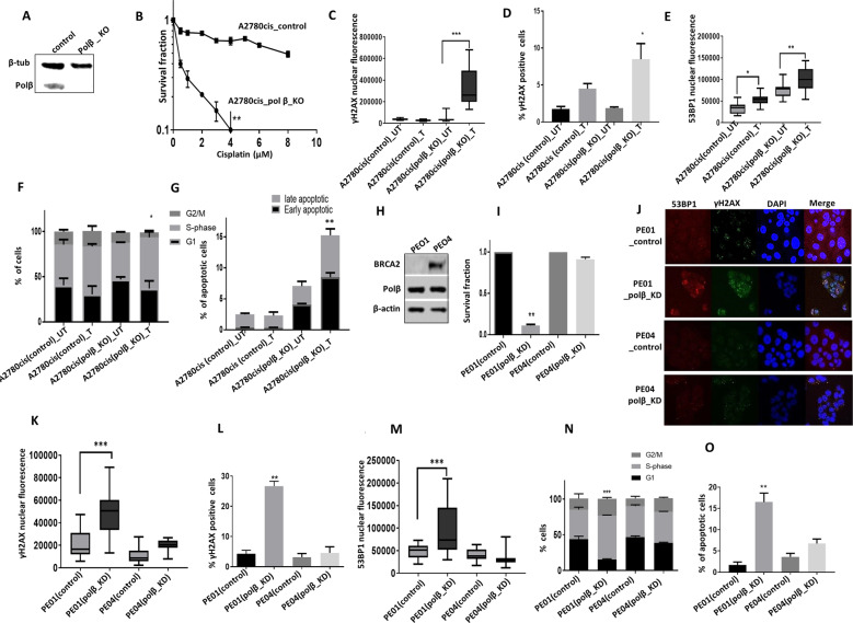
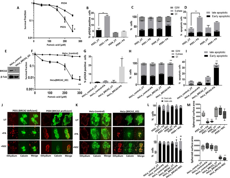
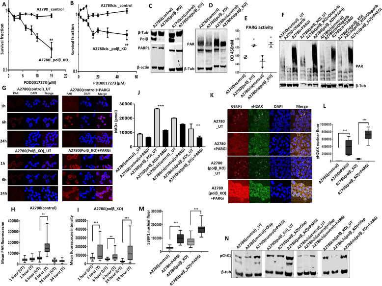
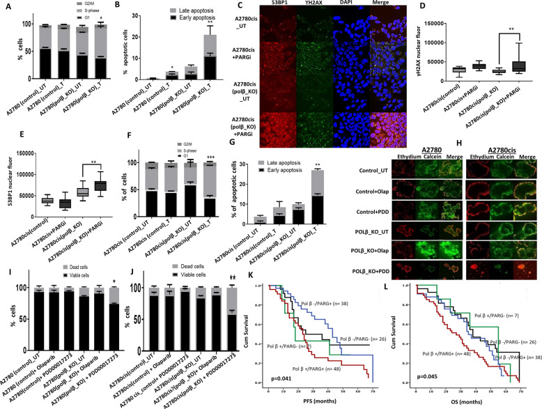
Targeting PARP1 [Poly(ADP-Ribose) Polymerase 1] for synthetic lethality is a new strategy for BRCA germ-line mutated or platinum sensitive ovarian cancers. However, not all patients respond due to intrinsic or acquired resistance to PARP1 inhibitor. Development of alternative synthetic lethality approaches is a high priority. DNA polymerase β (Polβ), a critical player in base excision repair (BER), interacts with PARP1 during DNA repair. Here we show that polβ deficiency is a predictor of platinum sensitivity in human ovarian tumours. Polβ depletion not only increased platinum sensitivity but also reduced invasion, migration and impaired EMT (epithelial to mesenchymal transition) of ovarian cancer cells. Polβ small molecular inhibitors (Pamoic acid and NSC666719) were selectively toxic to BRCA2 deficient cells and associated with double-strand breaks (DSB) accumulation, cell cycle arrest and increased apoptosis. Interestingly, PARG [Poly(ADP-Ribose) Glycohydrolase] inhibitor (PDD00017273) [but not PARP1 inhibitor (Olaparib)] was synthetically lethal in polβ deficient cells. Selective toxicity to PDD00017273 was associated with poly (ADP-ribose) accumulation, reduced nicotinamide adenine dinucleotide (NAD+) level, DSB accumulation, cell cycle arrest and increased apoptosis. In human tumours, polβ-PARG co-expression adversely impacted survival in patients. Our data provide evidence that polβ targeting is a novel strategy and warrants further pharmaceutical development in epithelial ovarian cancers.


Conflict of interest statement

The authors declare no competing interests.

Figures

Fig. 1
Fig. 1. High Polβ expression is linked to aggressive ovarian cancers.
A Immunohistochemical expression of Polβ in ovarian cancers. B Kaplan–Meier curve for overall survival in: whole cohort. C Kaplan–Meier curve for Polβ mRNA expression. D Cisplatin sensitivity by clonogenic survival assay in A2780 and A2780cis. E Polβ nuclear and cytoplasmic extracts in A2780 and A2780cis treated with 5 µM cisplatin. Lysates collected 48 h post treatment. F Polβ siRNA knock down in A2780cis cells. Cell cycle analysis for A2780cis control and Polβ_KD cells is shown here, G ATR and p-CHK1 protein expression in A2780cis Polβ_KD cells. H Quantification of ATR and p-CHK1 by western blot. I Cisplatin sensitivity by clonogenic survival assay in A2780cis cells control and A2780cis_Polβ_KD cells. J Quantification of γH2AX positive cells by flow cytometry in A2780cis cells control and Polβ_knock down treated with 5 µM cisplatin for 24 h. K Cell cycle analysis by flow cytometry in A2780cis cells control and Polβ_knockdown treated with 5 μM cisplatin. L AnnexinV analysis for apoptotic cells in A2780cis cells control and Polβ_knock down treated with 5 μM cisplatin. For Flow cytometry cells were seeded and transfected with scrambled control or Polβ siRNA. At day 3 controls and knockdown cells were re platted in six-well plates overnight and treated with 5 μM cisplatin and analyzed by flow cytometry on day 5. Transfection efficiency was confirmed by western blotting. Figures are representative of three or more independent experiments.
Fig. 2
Fig. 2. Polβ depletion in ovarian cancer cell lines.
A Polβ siRNA transfection in PEO4 cells. B Cisplatin sensitivity by clonogenic survival assay in PEO4 scrambled control transfected cells and PEO4 Polβ_knockdown. PEO4 scrambled control and PEO4_Polβ_knockdown cells were treated with 5 μM cisplatin and on day 5 cells were analyzed by flow cytometry. C Representative photomicrographic images for immunofluorescence staining of γH2AX and 53BP1 in PEO4 scrambled control and polβ knockdown cells treated with 5 μM cisplatin. D Quantification of γH2AX nuclear fluorescence by ImageJ software. E Quantification of γH2AX positive cells by flow cytometry. F Quantification of 53BP1 nuclear fluorescence by ImageJ software. G Cell cycle analysis by flow cytometry. H Annexin V analysis by flow cytometry. I Polβ siRNA transfection in A2780 cells and Cisplatin sensitivity by clonogenic survival assay in A2780 scrambled control and Polβ_Knockdown cells. J Polβ knock out by CRISPR-Cas9 in A2780 cells and Cisplatin sensitivity by clonogenic survival assay in A2780 control and Polβ_Knock out cells. K Quantification of γH2AX nuclear fluorescence by ImageJ software. A2780 control and A2780 Polβ_knock out cells were treated with 1 μM cisplatin and analyzed by Immunofluorescence or flow cytometry on day 5. L Quantification of γH2AX positive cells by flow cytometry. M Quantification of 53BP1 nuclear fluorescence by ImageJ software. N Cell cycle analysis by flow cytometry. O Annexin V analysis by flow cytometry. A2780 control and Polβ_KO cells were plated overnight and treated with 1 μM cisplatin for 24 h. After incubation, cells were collected and stained as per the flow cytometry protocol detailed in the methods. Figures are representative of three or more independent experiments.
Fig. 3
Fig. 3. Polβ knock out increases platinum sensitivity.
A Polβ CRISPR_knockout in A2780cis cells. B Cisplatin sensitivity in A2780cis control and Polβ-knockout cells. C Quantification of γH2AX nuclear fluorescence by ImageJ software. D Quantification of γH2AX positive cells by flow cytometry. E Quantification of 53BP1 nuclear fluorescence by imageJ software. F Cell cycle analysis by flow cytometry. G Annexin V analysis by flow cytometry. For A2780cis control and Polβ_knockout cells, Cells were treated with 5 μM cisplatin for 24 h and analyzed by flow cytometry or cells were plated on coverslips overnight and treated with 5 μM cisplatin for 24 h. Cells were then fixed and stained for immunofluorescence as detailed in the methods. All figures are representative of three or more experiments. H BRCA2 and polβ expression by western blot in PEO1 and PEO4 cells. I Survival fraction in PEO1 and PEO4 control and Polβ_knockdown cells. J Representative photomicrographic images of PEO1 and PEO4 control and Polβ_knockdown cells. K Quantification of γH2AX nuclear fluorescence by ImageJ software. L γH2AX positive cells analysis by flow cytometry. M Quantification of 53BP1 nuclear fluorescence by ImageJ software. N Cell cycle analysis by flow cytometry. O Annexin V analysis by flow cytometry. PEO1 and PEO4 control and Polβ_knockdown cells were transfected with Polβ SiRNA; on day 4 cells were plated in six-well plates overnight and analyzed by flow cytometry on day 5. Figures are representative of three or more experiments.
Fig. 4
Fig. 4. Polβ inhibitors induce synthetic lethality in BRCA2 deficient ovarian cancer cells.
A Pamoic acid sensitivity by clonogenic survival assay in PEO1 and PEO4 cells. B γH2AX analysis by flow cytometry for PEO1 and PEO4 cells treated with Pamoic acid (250 μM). C Cell cycle analysis by flow cytometry in PEO1 and PEO4 cells treated with Pamoic acid (250 μM) for 24 h. D Annexiγn V analysis by flow cytometry in PEO1 and PEO4 cells treated with Pamoic acid (250 μM) for 24 h. E BRCA2 western blot in HeLa control and HeLa BRCA2_knockdown cells. F Pamoic acid sensitivity in HeLa control and HeLa BRCA2_knock down cells by clonogenic survival assay is shown here. γH2AX analysis by flow cytometry (G), cell cycle analysis by flow cytometry (H) and annexinV analysis by flow cytometry (I) in HeLa control and HeLa BRCA2_knockdown cells treated with Pamoic acid (250 μM) for 24 h. Representative photomicrographic images of Pamoic acid (250 μM) and NSC666719 (250 μM) treated 3D-spheres of: PEO1 and PEO4 cells (J), HeLa control and HeLa (BRCA2_KD) (K). L Quantification of spheroids cell viability by flow cytometry. M Quantification of spheroids size by ImageJ. Figures are representative of three or more experiments. Error bars represent standard deviation between experiments.
Fig. 5
Fig. 5. Polβ knock out cells is sensitive to PARG inhibitor.
A PARG inhibitor sensitivity by clonogenic survival assay in A2780 control and Polβ_knock out cells. B PARG inhibitor sensitivity by clonogenic survival assay in A2780cis control and Polβ_knock out cells. C Polβ, PARP1 levels by western blot in A2780 and A2780cis control and Polβ_knockout cells. D Poly (ADP) ribose polymers levels in A2780 and A2780cis control and Polβ_knock out cells. E PARG ELISA assay in A2780 control, A2780 (Polβ_KO), A2780cis control and A2780cis (Polβ_KO) cells. F Poly (ADP) ribose polymers levels in A2780 and A2780cis control and Polβ_knock out cells treated with Olaparib or PARG inhibitor. Cells were treated with Olaparib (10 μM) or PARGi (20 μM) for 16 h then extracted for western blot. G Immunofluorescence staining for poly (ADP) ribose polymers in A2780 control and Polβ_knockout cells untreated or treated with PARGi (20 μM) for 1, 6, and 24 h. H, I Quantification of Poly (ADP) ribose polymers fluorescence by imageJ software. J Quantification of NAD+ levels in A2780 and A2780cis control and Polβ_knock out cells. K Representative photomicrographic images of 53BP1 and γH2AX immunofluorescence in A2780 and A2780cis control and Polβ_knock out cells treated with PARGi (20 μM) for 16 h. L Quantification of γH2AX nuclear fluorescence by ImageJ software. M Quantification of 53BP1 nuclear fluorescence by ImageJ software. N p-CHK1 levels by western blot in A2780 and A2780cis control and Polβ_knock out treated with Olaparib (10 μM) or PARGi (20 μM) for 16 h. Figures are representative of three or more experiments. Error bars represent standard deviation between experiments.
Fig. 6
Fig. 6. Mechanisitc studies of PARG inhibitor in ovarian cancer.
A Cell cycle analysis by flow cytometry. B Annexin V analysis by flow cytometry for A2780 cells control and Polβ_knockout cells treated with 20 μM PARGi for 24 h. C Representative photomicrographic images for 53BP1 and γH2AX immunofluorescence staining in A2780 control and Polβ_knockout cells treated with 20 μM PARGi. D Quantification by γH2AX nuclear fluorescence by ImageJ software. E Quantification of 53BP1 nuclear fluorescence by ImageJ software. F Cell cycle analysis by flow cytometry. G Annexin V analysis by flow cytometry. A2780cis cells control and Polβ_knockout cells were treated with 20 μM PARGi for 24 h cells were collected and analysed as per the flow cytometry protocol or cells were seeded on coverslips and treated with PARGi(20 μM) for 24 h before staining for immunofluorescence protocol. H Representative photomicrographic images for A2780 control and Polβ_knockout spheroids treated with Olaparib (10 μM) or PARGi (20 μM). I, J Quantification of spheroids cell viability is shown here. Figures are representative of three or more independent experiments. K Polβ and PARG co-expression and Kaplan–Meier curve for ovarian cancer progression-free survival (PFS). L Polβ and PARG co-expression and Kaplan–Meier curve for overall survival in ovarian cancer.

Similar articles

Cited by

References

    1. Moore K, Colombo N, Scambia G, Kim BG, Oaknin A, Friedlander M, et al. Maintenance olaparib in patients with newly diagnosed advanced ovarian cancer. N Engl J Med. 2018;379:2495–505. - PubMed
    1. Mirza MR, Monk BJ, Herrstedt J, Oza AM, Mahner S, Redondo A, et al. Niraparib maintenance therapy in platinum-sensitive, recurrent ovarian cancer. N. Engl J Med. 2016;375:2154–64. - PubMed
    1. Swisher EM, Lin KK, Oza AM, Scott CL, Giordano H, Sun J, et al. Rucaparib in relapsed, platinum-sensitive high-grade ovarian carcinoma (ARIEL2 Part 1): an international, multicentre, open-label, phase 2 trial. Lancet Oncol. 2017;18:75–87. - PubMed
    1. D’Andrea AD. Mechanisms of PARP inhibitor sensitivity and resistance. DNA Repair. 2018;7:172–6. - PubMed
    1. Lindahl T. Repair of intrinsic DNA lesions. Mutat Res. 1990;238:305–11. - PubMed

Publication types

LinkOut - more resources