. 2021 Mar 5;4(1):299.
doi: 10.1038/s42003-021-01818-7.

Engineering subtilisin proteases that specifically degrade active RAS

Affiliations

Engineering subtilisin proteases that specifically degrade active RAS

Yingwei Chen et al. Commun Biol. .

Abstract

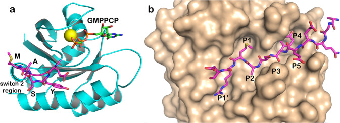
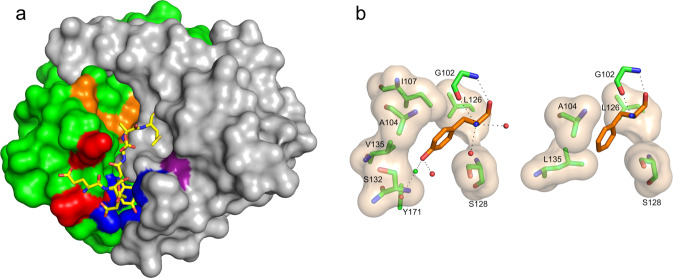
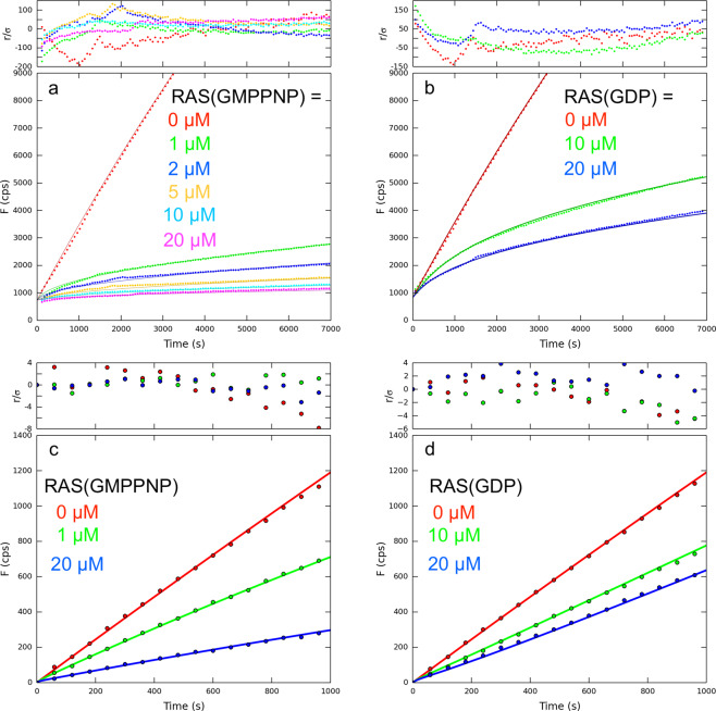
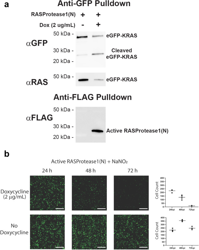
We describe the design, kinetic properties, and structures of engineered subtilisin proteases that degrade the active form of RAS by cleaving a conserved sequence in switch 2. RAS is a signaling protein that, when mutated, drives a third of human cancers. To generate high specificity for the RAS target sequence, the active site was modified to be dependent on a cofactor (imidazole or nitrite) and protease sub-sites were engineered to create a linkage between substrate and cofactor binding. Selective proteolysis of active RAS arises from a 2-step process wherein sub-site interactions promote productive binding of the cofactor, enabling cleavage. Proteases engineered in this way specifically cleave active RAS in vitro, deplete the level of RAS in a bacterial reporter system, and also degrade RAS in human cell culture. Although these proteases target active RAS, the underlying design principles are fundamental and will be adaptable to many target proteins.


Conflict of interest statement

The authors declare no competing interests.

Figures

Fig. 1
Fig. 1. Target sequence (QEEYSAM) in RAS and in the active site of a RAS-specific protease.
a Structure of RAS with a bound GTP analog (PDB code 6Q21),, highlighting the YSAM site in Switch 2. b RAS-specific protease based on an X-ray structure of 3BGO.pdb. Cognate sequence QEEYSAM-RD is modeled in the binding cleft. Substrate residues are denoted P1 through P5, numbering from the scissile bond toward the N-terminus of the substrate. The substrate amino acid on the leaving group side is denoted P1’.
Fig. 2
Fig. 2. kcat/KM as a function of mutation in the P4 pocket.
a Protease1(N) variants in 1 mM nitrite. b Protease1(I) variants in 10 mM imidazole. c Comparison of P4 specificity for Protease2(N) in 1 mM nitrite and Protease2(I) in 10 mM imidazole for the five highest activity sDXKAM-AMC substrates.
Fig. 3
Fig. 3. kcat/KM for the target substrate QEEYSAM-AMC as a function of cofactor.
a Protease2(N) and RASProtease(N) vs. nitrite concentration. b Protease2(I) and RASProtease(I) vs. imidazole concentration. The activity of a progenitor protease (SBT160) with a complete catalytic triad (D32, H64, S221) is shown for comparison.
Fig. 4
Fig. 4. Conformational and chemical rescue by native and near-native sequences.
a kcat/KM as a function of nitrite concentration for RASProtease(N) with the substrates QEEYSAM-AMC and QEEISAM-AMC. (b) kcat/KM as a function of imidazole concentration for RASProtease(I) with the substrates QEEYSAM-AMC and QEEISAM-AMC.
Fig. 5
Fig. 5. Engineering a protease directed against active RAS.
a Surface representation of RASProtease(I) with substrate binding sites colored purple (S1), orange (S2 and catalytic residues), red (S3), and blue (S4) with the bound YSAM peptide overlaid. b Interactions at the P4 site of RASProtease(I) (left) and its progenitor Protease1(N) (right). The van der Waals interactions between residues in the protease active site (carbons colored green) and the bound P4 residue (carbons colored orange) are represented as a tan surface. Hydrogen bonds are represented as black dashed lines.
Fig. 6
Fig. 6. Kinetics of AMC release from QEEYSAM-AMC by 100 nM RASProtease in the presence of RAS.
a RASProtease(I) + RAS(GMPPNP). b RASProtease(I) + RAS(GDP). (a) and (b) were measured in the presence of 1 µM QEEYSAM-AMC and 1 mM imidazole. c RASProtease(N) + RAS(GMPPNP). d RASProtease(N) + RAS(GDP). (c) and (d) were measured in the presence of 1 µM QEEYSAM-AMC and 1 mM nitrite. Data points are solid circles. Global fit to mechanism 1 are solid lines. Residuals are plotted above each graph.
Fig. 7
Fig. 7. RASProtease(I) with 100 µM imidazole added to the culture media at 17 h.
Intact RAS fusion protein is 35,974 daltons. The N- and C- terminal fragments of RAS are 13,579 (r2) and 22,413 daltons (r1), respectively. The C-terminal fragment of the I-domain is 6,175 daltons (i1). Markers: 250, 150, 100, 75, 50, 37, 25, 20, 15, 10 kDa.
Fig. 8
Fig. 8. RAS-specific protease activity in cells.
a Western blot analysis of cells co-transfected with eGFP-KRAS and RASProtease shows the appearance of a KRAS cleavage product upon induction of the active protease when probed with an anti-GFP antibody following a GFP pull-down. Sodium nitrite was added to the cell culture medium at a final concentration of 1 mM to mitigate potential variability in cellular nitrite concentrations. Appearance of this product coincides with depletion of a RAS-reactive band when probed with an anti-RAS antibody. Appearance of cleaved eGFP-KRAS also coincides with expression of activated protease that has cleaved its inhibitory I-domain. b Induction of the active protease in HEK 293 T cells at 24 h after transfection with nitrite supplemented culture medium results in a marked decrease in GFP fluorescence at 48 and 72 h after transfection compared to the same cells without induction of protease expression. The scale bar is 200 µm.
Fig. 9
Fig. 9. Two views of the domain structure for RAS-specific subtilisin (6UAO.pdb) with substrate bound.
The N-terminal domain (N) is green, C-terminal domain (C) is gray, and QEEYSAM substrate (S) is yellow.

Similar articles

Cited by

References

    1. Vidimar V, et al. An engineered chimeric toxin that cleaves activated mutant and wild-type RAS inhibits tumor growth. Proc. Natl Acad. Sci. USA. 2020;117:16938–16948. doi: 10.1073/pnas.2000312117. - DOI - PMC - PubMed
    1. Huang L, Hofer F, Martin GS, Kim SH. Structural basis for the interaction of Ras with RalGDS. Nat. Struct. Biol. 1998;5:422–426. doi: 10.1038/nsb0698-422. - DOI - PubMed
    1. Buhrman G, Kumar VS, Cirit M, Haugh JM, Mattos C. Allosteric modulation of Ras-GTP is linked to signal transduction through RAF kinase. J. Biol. Chem. 2011;286:3323–3331. doi: 10.1074/jbc.M110.193854. - DOI - PMC - PubMed
    1. Lu S, et al. Ras conformational ensembles, allostery, and signaling. Chem. Rev. 2016;116:6607–6665. doi: 10.1021/acs.chemrev.5b00542. - DOI - PubMed
    1. Milburn MV, et al. Molecular switch for signal transduction: structural differences between active and inactive forms of protooncogenic ras proteins. Science. 1990;247:939–945. doi: 10.1126/science.2406906. - DOI - PubMed

Publication types