. 2021 Jun 25;38(7):2715-2731.
doi: 10.1093/molbev/msab060.

Identifying Primate ACE2 Variants That Confer Resistance to SARS-CoV-2

Affiliations

Identifying Primate ACE2 Variants That Confer Resistance to SARS-CoV-2

Maloyjo Joyraj Bhattacharjee et al. Mol Biol Evol. .

Abstract

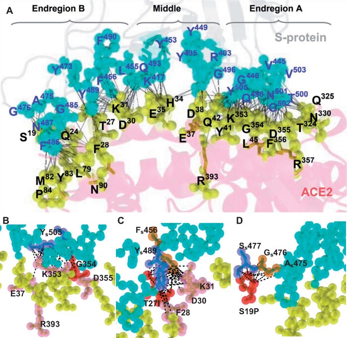
SARS-CoV-2 infects humans through the binding of viral S-protein (spike protein) to human angiotensin I converting enzyme 2 (ACE2). The structure of the ACE2-S-protein complex has been deciphered and we focused on the 27 ACE2 residues that bind to S-protein. From human sequence databases, we identified nine ACE2 variants at ACE2-S-protein binding sites. We used both experimental assays and protein structure analysis to evaluate the effect of each variant on the binding affinity of ACE2 to S-protein. We found one variant causing complete binding disruption, two and three variants, respectively, strongly and mildly reducing the binding affinity, and two variants strongly enhancing the binding affinity. We then collected the ACE2 gene sequences from 57 nonhuman primates. Among the 6 apes and 20 Old World monkeys (OWMs) studied, we found no new variants. In contrast, all 11 New World monkeys (NWMs) studied share four variants each causing a strong reduction in binding affinity, the Philippine tarsier also possesses three such variants, and 18 of the 19 prosimian species studied share one variant causing a strong reduction in binding affinity. Moreover, one OWM and three prosimian variants increased binding affinity by >50%. Based on these findings, we proposed that the common ancestor of primates was strongly resistant to and that of NWMs was completely resistant to SARS-CoV-2 and so is the Philippine tarsier, whereas apes and OWMs, like most humans, are susceptible. This study increases our understanding of the differences in susceptibility to SARS-CoV-2 infection among primates.

Keywords: : COVID-19; ACE2; S-protein; resistant to SARS-CoV-2.


Figures

Fig. 1.
Fig. 1.
Cladogram of the primates studied and changes at key ACE2 residues during primate evolution. The inferred amino acids at the key ACE2 residues in the common ancestor of primates are shown at the root of the cladogram. The nine human ACE2 variants identified in this study are shown at the bottom of the figure, whereas the nonhuman primate variants are shown on the tree branches. The effect of each variant on binding affinity was evaluated by RBD attachment assay (see fig. 3) and is indicated by a color according to the color code at the bottom of this figure. Although site 92 is not a key binding residue, it is close to the N90 glycosylation site and the T92I mutation we identified in humans was found by Chan et al. (2020) to increase the binding affinity of ACE2 to S-protein; however, as its effect on binding affinity was not evaluated in this study, it is marked by “?” The T27A substitution in rhesus macaque (Macaca mulatta) is marked by an * because it only appeared in one individual we collected (supplementary data 4, Supplementary Material online).
Fig. 2.
Fig. 2.
The interaction between RBD and ACE2 variants measured by the RBD attachment assay. (A) Schematic illustration of the RBD attachment assay. Complementation of an active NanoLuc is achieved when the LgBiT and Sm-BiT are brought together upon interaction of RBD and ACE2. (B) HeLa cells transfected with mock construct or SmBiT-ACE2 were treated with RBD-LgBiT and subjected to bioluminescence detection for 1 h. Peak bioluminescence signal was detected at 10 min after the addition of RBD-LgBiT. (C) FLS was mixed with RBD-LgBiT at different molar ratios before incubation with HeLa cells transfected with SmBiT-ACE2. Bioluminescence signal was reduced by increasing the molar ratio of FLS. (D) Expression levels of the wild-type and human ACE2 variants were confirmed by western blotting. Two separate clones were tested for each variant transfection. The GAPDH expression served as an internal control for each experiment.
Fig. 3.
Fig. 3.
Measuring RBD attachment on primate ACE2 variants. (A) RBD attachment activity measured on 9 human and 27 nonhuman primate ACE2 variants. The maximum bioluminescence signal measured in the RBD attachment assay for each variant was normalized by that of wild-type ACE2. Data are mean ± SEM of at least three replicates. (B) Intracellular protein expression levels of 27 single primate ACE2 variants, 2 double mutants, and 1 triple mutant were confirmed by western blotting. Expressions of two independent constructs for each variant were shown.
Fig. 4.
Fig. 4.
Binding of S1-hFc to cell surface ACE2 variants. (A) Schematic illustration of the binding assay. HeLa cells transfected with ACE2 expression constructs were incubated 1 h with recombinant S1-hFc and cell surface bound S1-hFc was detected by immunofluorescence staining. (B) Representative images of S1-hFc (red) and HeLa cells expressing human ACE2 variants (green). (C) Representative images of S1-hFc (red) and HeLa cells expressing nonhuman primate ACE2 variants (green).
Fig. 5.
Fig. 5.
The binding interface and atomic contacts between ACE2 and S-protein. (A) Binding between S-protein and ACE2 (in cyan and yellow spheres, respectively). The ACE2 binding residues (in yellow spheres) on the interface are classified into Endregion A, Middle, and Endregion B. Each contact between an ACE2 atom and an S-protein atom is analytically calculated and represented by a black dotted line. K353 of ACE2 has 42 atomic contacts, the strongest binding residue on the ACE2–S-protein interface. The highest density of atomic contacts between ACE2 and S-protein is detected at K353, Y41, and K31 of ACE2. (BD) Close-up views of atomic contacts on the interface. (B) G354 in Endregion A is situated in the middle of the surface patch of K353, G354, and D355 (in red spheres), which strongly binds to the most crucial residue Ys505 (in slate) of S-protein. The amide backbone of G354 is replaced by a bulky polar side chain in G354Q (in red), leading to the removal of all five atomic contacts to Ys505 and reducing the binding affinity of K353, R393, and E37 (in pink) to Ys505. (C) The aromatic rings of Ys489 (in slate) and Fs456 (in orange) are docked into the groove of I27, F28, D30, and K31 (in pink). The polar side chain of T27 is replaced by a hydrophobic side-chain in the T27I mutation (in red), adding 3 and 5 atomic contacts to Ys489 and Fs456, respectively. (D) The S19P mutation on Endregion B gains 6 and 3 atomic contacts to Ss477 and Gs476, respectively, conferring a large increase in binding affinity to S-protein.
Fig. 6.
Fig. 6.
Electrostatic potential analysis of the binding interface of ACE2 for the wild-type Q42 and mutants E42 and L42. The atoms on the binding interface of ACE2 are colored according to their charges. The ACE binding interface displays charged surfaces with iso-surfaces drawn at 1.0 (blue) and −1.0 (red) kT/e, where k, T, and e denote the Boltzmann constant, the temperature, and the charge units, respectively. (A) ACE2 and S-protein display two distinctive patterns of electrostatic surfaces. (B) ACE2 is rotated to show that its binding interface includes mostly negatively charged areas (red). (C) After rotation, the S-protein interface shows mostly hydrophobic areas (white) with negatively charged patches (red). As indicated, Q498 and Y449 (in pink dots that represent atoms) are situated on the hydrophobic surface, interacting with Q42 of ACE2. (D) The wild-type Q42 (in pink dots) of ACE2 exhibits a mildly negatively charged surface. In comparison, the Q42E (in pink dots) mutation enhances the negatively charged surface with its neighboring residues (E), which may largely reject the interaction of ACE2 with S-protein, whereas the Q42L (in pink dots) mutation expands into a larger hydrophobic surface (F), strongly promoting the interaction of ACE2 with the hydrophobic surface of S-protein in (C).
Fig. 7.
Fig. 7.
The effects of the M82I, M82T, and M82S mutations on the binding interface between ACE2 and S-protein. The shortest distance is 2.44 Å between M82 of ACE2 and Fs486 of S-protein and 5.85 Å between Y83 of ACE2 and Fs486 of S-protein. (A) The phenyl ring of Fs486 is oriented into the groove between M82 and Y83 of human wild-type ACE2 (PDB6m17). Pairwise atomic distances are displayed as dashed lines in yellow. (B) The residue packing at the local region of the M82I-mutant ACE2 is significantly altered. A prominent modification is that the phenyl ring of Fs486 is reoriented and situated away from the hydroxyphenyl ring of Y83 of the M82I mutant. (CD) The replacement of the bulky hydrophobic methyl side chain in M82 by a polar side chain in T82 (C) or S82 (D) severely inhibits direct atomic contacts with the phenyl ring of Fs486, weakening the interaction between Fs486 and Y83 of ACE2 in M82T and M82S.

References

    1. The 1000 Genome Project Consortium. 2015. A global reference for human genetic variation. Nature 526:68–74. - PMC - PubMed
    1. Baker NA, Sept D, Joseph S, Holst MJ, McCammon JA.. 2001. Electrostatics of nanosystems: application to microtubules and the ribosome. Proc Natl Acad Sci U S A. 98(18):10037–10041. - PMC - PubMed
    1. Bergström A, McCarthy SA, Hui R, Almarri MA, Ayub Q, Danecek P, Chen Y, Felkel S, Hallast P, Kamm J, et al.2020. Insights into human genetic variation and population history from 929 diverse genomes. Science 367(6484):eaay5012. - PMC - PubMed
    1. Camacho C, Coulouris G, Avagyan V, Ma N, Papadopoulos J, Bealer K, Madden TL.. 2009. BLAST+: architecture and applications. BMC Bioinformatics 10(1):421–429. - PMC - PubMed
    1. Cao Y, Li L, Feng Z, Wan S, Huang P, Sun X, Wen F, Huang X, Ning G, Wang W.. 2020. Comparative genetic analysis of the novel coronavirus (2019-nCoV/SARS-CoV-2) receptor ACE2 in different populations. Cell Discov. 6(1):1–4. - PMC - PubMed

Publication types