

Increase in Ventricle Size and the Evolution of White Matter Changes on Serial Imaging in Critically Ill Patients with COVID-19
- PMID: 33674942
- PMCID: PMC7935478
- DOI: 10.1007/s12028-021-01207-2
Increase in Ventricle Size and the Evolution of White Matter Changes on Serial Imaging in Critically Ill Patients with COVID-19
Abstract
Background: Evolution of brain magnetic resonance imaging (MRI) findings in critically ill patients with coronavirus disease 2019 (COVID-19) is unknown.
Methods: We retrospectively reviewed 4530 critically ill patients with COVID-19 admitted to three tertiary care hospitals in New York City from March 1 to June 30, 2020 to identify patients who had more than one brain MRI. We reviewed the initial and final MRI for each patient to (1) measure the percent change in the bicaudate index and third ventricular diameter and (2) evaluate changes in the presence and severity of white matter changes.
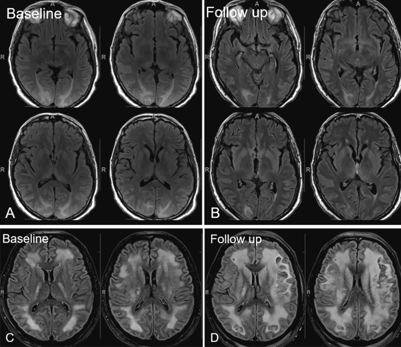
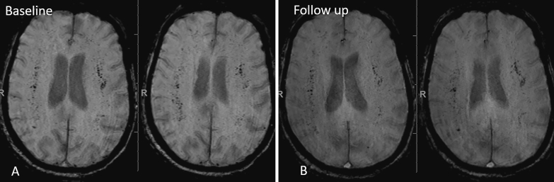
Results: Twenty-one patients had two MRIs separated by a median of 22 [Interquartile range (IQR) 14-30] days. Ventricle size increased for 15 patients (71%) between scans [median bicaudate index 0.16 (IQR 0.126-0.181) initially and 0.167 (IQR 0.138-0.203) on final imaging (p < 0.001); median third ventricular diameter 6.9 mm (IQR 5.4-10.3) initially and 7.2 mm (IQR 6.4-10.8) on final imaging (p < 0.001)]. Every patient had white matter changes on the initial and final MRI; between images, they worsened for seven patients (33%) and improved for three (14%).
Conclusions: On serial imaging of critically ill patients with COVID-19, ventricle size frequently increased over several weeks. White matter changes were often unchanged, but in some cases they worsened or improved, demonstrating there is likely a spectrum of pathophysiological processes responsible for these changes.
Keywords: COVID-19; Leukoencephalopathy; SARS-CoV-2; Ventricle.
© 2021. Springer Science+Business Media, LLC, part of Springer Nature and Neurocritical Care Society.
Conflict of interest statement
All authors report no disclosures.
Figures
Similar articles
-
Serial Imaging of Virus-Associated Necrotizing Disseminated Acute Leukoencephalopathy (VANDAL) in COVID-19.AJNR Am J Neuroradiol. 2021 Jan;42(2):279-284. doi: 10.3174/ajnr.A6898. Epub 2020 Oct 22. AJNR Am J Neuroradiol. 2021. PMID: 33093131 Free PMC article.
-
Cerebral Microbleeds and Leukoencephalopathy in Critically Ill Patients With COVID-19.Stroke. 2020 Sep;51(9):2649-2655. doi: 10.1161/STROKEAHA.120.030940. Epub 2020 Aug 5. Stroke. 2020. PMID: 32755456 Free PMC article.
-
Baseline Characteristics and Outcomes of 1591 Patients Infected With SARS-CoV-2 Admitted to ICUs of the Lombardy Region, Italy.JAMA. 2020 Apr 28;323(16):1574-1581. doi: 10.1001/jama.2020.5394. JAMA. 2020. PMID: 32250385 Free PMC article.
-
Clinical, Imaging, and Lab Correlates of Severe COVID-19 Leukoencephalopathy.AJNR Am J Neuroradiol. 2021 Apr;42(4):632-638. doi: 10.3174/ajnr.A6966. Epub 2021 Jan 7. AJNR Am J Neuroradiol. 2021. PMID: 33414226 Free PMC article.
-
Brain abnormalities in COVID-19 acute/subacute phase: A rapid systematic review.Brain Behav Immun. 2020 Oct;89:543-554. doi: 10.1016/j.bbi.2020.07.014. Epub 2020 Jul 17. Brain Behav Immun. 2020. PMID: 32682993 Free PMC article.
Cited by
-
Imaging Markers of Neurologic Damage in COVID-19: A Systematic Review.Curr Med Chem. 2023;30(9):1086-1106. doi: 10.2174/0929867329666220701124945. Curr Med Chem. 2023. PMID: 35786328
-
Acute and long-term effects of COVID-19 on brain and mental health: A narrative review.Brain Behav Immun. 2025 Jan;123:928-945. doi: 10.1016/j.bbi.2024.11.007. Epub 2024 Nov 3. Brain Behav Immun. 2025. PMID: 39500417 Review.
-
Brain MRI findings in severe COVID-19 patients: a meta-analysis.Front Neurol. 2023 Oct 12;14:1258352. doi: 10.3389/fneur.2023.1258352. eCollection 2023. Front Neurol. 2023. PMID: 37900601 Free PMC article.
-
Are the Post-COVID-19 Posttraumatic Stress Disorder (PTSD) Symptoms Justified by the Effects of COVID-19 on Brain Structure? A Systematic Review.J Pers Med. 2023 Jul 15;13(7):1140. doi: 10.3390/jpm13071140. J Pers Med. 2023. PMID: 37511753 Free PMC article. Review.
-
Topographical Distribution of Neuroanatomical Abnormalities Following COVID-19 Invasion : A Systematic Literature Review.Clin Neuroradiol. 2024 Mar;34(1):13-31. doi: 10.1007/s00062-023-01344-5. Epub 2023 Sep 11. Clin Neuroradiol. 2024. PMID: 37697012 Free PMC article.
References
-
- Chougar L, Shor N, Weiss N, Galanaud D, Leclercq D, Mathon B, et al. Retrospective observational study of brain magnetic resonance imaging findings in patients with acute SARS-CoV-2 infection and neurological manifestations. Radiology. 2020;297:E313–E323. doi: 10.1148/radiol.2020202422. - DOI - PMC - PubMed
MeSH terms
LinkOut - more resources
Full Text Sources
Other Literature Sources
Medical
Miscellaneous
