. 2021 Mar;35(3):e21412.
doi: 10.1096/fj.201903203RR.

A cell-penetrating CD40-TRAF2,3 blocking peptide diminishes inflammation and neuronal loss after ischemia/reperfusion

Affiliations

A cell-penetrating CD40-TRAF2,3 blocking peptide diminishes inflammation and neuronal loss after ischemia/reperfusion

Jose-Andres C Portillo et al. FASEB J. 2021 Mar.

Abstract

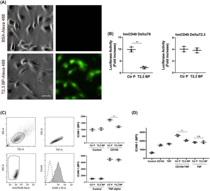
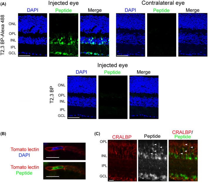
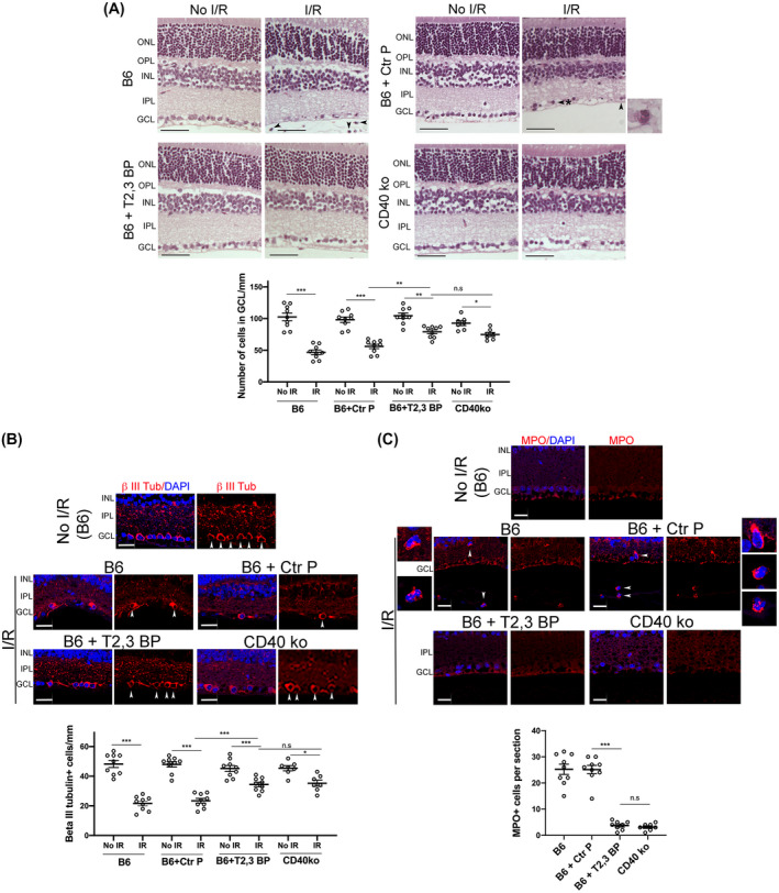
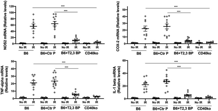
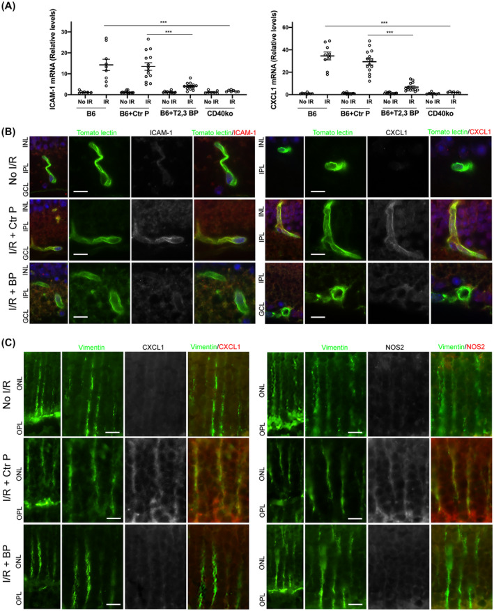
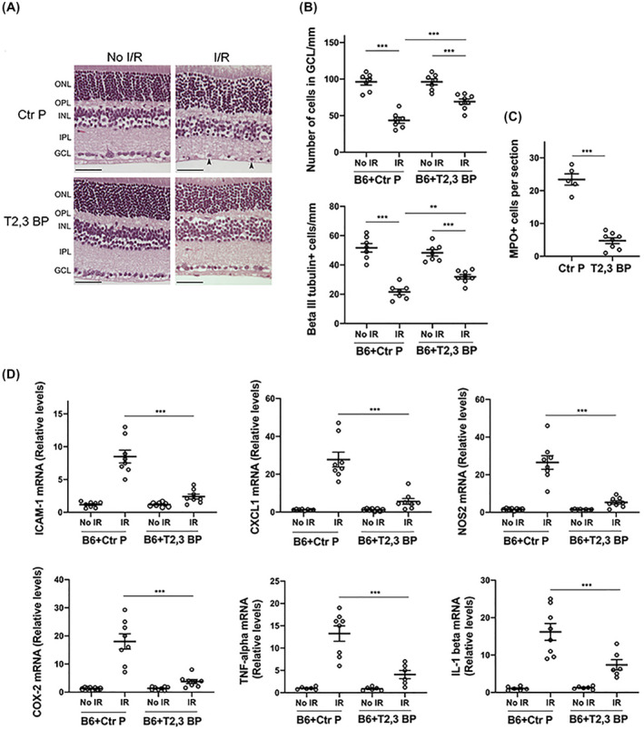
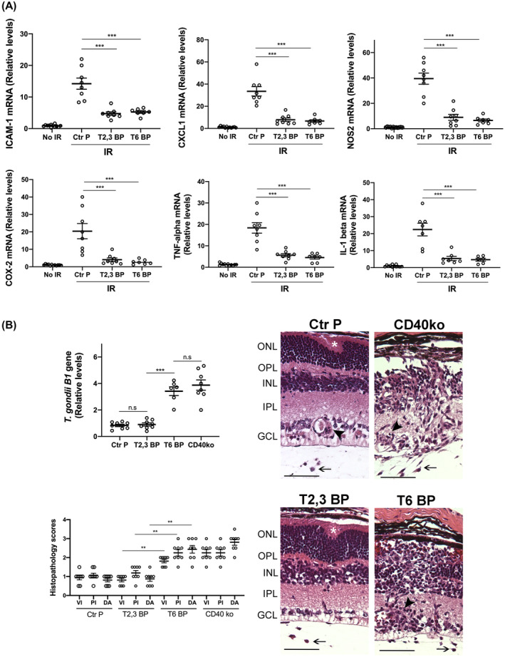
While the administration of anti-CD154 mAbs in mice validated the CD40-CD154 pathway as a target against inflammatory disorders, this approach caused thromboembolism in humans (unrelated to CD40 inhibition) and is expected to predispose to opportunistic infections. There is a need for alternative approaches to inhibit CD40 that avoid these complications. CD40 signals through TRAF2,3 and TRAF6-binding sites. Given that CD40-TRAF6 is the pathway that stimulates responses key for cell-mediated immunity against opportunistic pathogens, we examined the effects of pharmacologic inhibition of CD40-TRAF2,3 signaling. We used a model of ischemia/reperfusion (I/R)-induced retinopathy, a CD40-driven inflammatory disorder. Intravitreal administration of a cell-penetrating CD40-TRAF2,3 blocking peptide impaired ICAM-1 upregulation in retinal endothelial cells and CXCL1 upregulation in endothelial and Müller cells. The peptide reduced leukocyte infiltration, upregulation of NOS2/COX-2/TNF-α/IL-1β, and ameliorated neuronal loss, effects that mimic those observed after I/R in Cd40-/- mice. While a cell-penetrating CD40-TRAF6 blocking peptide also diminished I/R-induced inflammation, this peptide (but not the CD40-TRAF2,3 blocking peptide) impaired control of the opportunistic pathogen Toxoplasma gondii in the retina. Thus, inhibition of the CD40-TRAF2,3 pathway is a novel and potent approach to reduce CD40-induced inflammation, while likely diminishing the risk of opportunistic infections that would otherwise accompany CD40 inhibition.

Keywords: endothelial; muller cell; polymorphonuclear leukocyte; retina; toxoplasma.


Conflict of interest statement

Drs. Carlos S. Subauste and M. Cecilia Subauste have a patent on the CD40‐TRAF2,3 blocking peptide. The other authors have no conflicts of interest to declare.

Figures

FIGURE 1
FIGURE 1
ri CD40‐TRAF2,3 blocking peptide penetrates cells, inhibits CD40‐TRAF2,3 signaling and impairs CD40‐driven ICAM‐1 upregulation. A, Human Müller cells were incubated in medium containing Alexa Fluor 488‐conjugated ri CD40‐TRAF2,3 blocking peptide (T2,3 BP) or Alexa Fluor 488‐conjugated bovine serum albumin (BSA; both at 10 µM) for 3 hours. Scale bar, 50 μm. Original magnification 400×. Images represent fluorescence of unfixed Müller cells after extensive washing of monolayers. B, Mouse endothelial cells (mHEVc) that express an NF‐κB response element that drives the transcription of a luciferase reporter plus either hmCD40 ΔT2,3 or hmCD40 ΔT6 were pre‐incubated with ri control peptide (Ctr P) or ri CD40‐TRAF2,3 blocking peptide (T2,3 BP; both at 1 µM) or medium alone followed by stimulation with human CD154. Data are expressed as fold increase in normalized luciferase activity in cells stimulated with CD154 compared to cells treated with respective peptide in the absence of CD154. C, Human retinal endothelial cells were treated with ri control peptide (Ctr P) or ri CD40‐TRAF2,3 blocking peptide (T2,3 BP; both at 1 µM) followed by stimulation with CD154 or TNF‐α (100 pg/mL) for 24 hours. Expression of ICAM‐1 was assessed by flow cytometry. Dot plot and histogram show gating strategy. ICAM‐1 was analyzed on live cells that did not stain with Aqua LIVE/DEAD kit. D. Human retinal endothelial cells were incubated with or without TNF‐α (30 pg/mL) followed by treatment with peptides and stimulation with CD154. Data shown represent mean ± SD of triplicate samples. Results are representative of three independent experiments. **< .01 by ANOVA
FIGURE 2
FIGURE 2
ri CD40‐TRAF2,3 blocking peptide penetrates retinal cells. A, B6 mice received Alexa Fluor 488‐conjugated ri CD40‐TRAF2,3 blocking peptide or non‐fluorescent ri CD40‐TRAF2,3 blocking peptide (both 1 μg) via intravitreal injection of one eye. Injected and contralateral eyes were collected after 48 hours and frozen sections were examined. GCL = Ganglion cell layer; IPL = Inner plexiform layer; INL = Inner nuclear layer. OPL = Outer plexiform layer; ONL = Outer nuclear layer. Scale bar, 50 µm. B, C, Retinas from mice injected with Alexa Fluor 488‐conjugated ri CD40‐TRAF2,3 blocking peptide were stained with DyLight 594 tomato lectin (labels neural endothelial cells, B) or with anti‐CRALBP antibody (labels Müller cells, C). Green fluorescence was detected in cytoplasmic processes that co‐stain with CRALBP (arrowheads). Scale bar 10 µm. Original magnification 600× for panel B and 400× for panel C, Results are representative of three independent experiments
FIGURE 3
FIGURE 3
ri CD40‐TRAF2,3 blocking peptide protects against cell loss in the GCL and infiltration by MPO+ leukocytes in retinas subjected to I/R. One eye from each B6 and Cd40−/− mouse was subjected to I/R. Contralateral non‐ischemic eye was used as control. Eyes subjected to I/R in B6 mice were treated intravitreously with or without ri control peptide (Ctr P), ri CD40‐TRAF2,3 blocking peptide (T2,3 BP; both 1 μg) 1 hour prior to an increase in IOP. Eyes were collected 2 days after I/R. A, Cell loss in the GCL is observed in ischemic eyes from B6 mice treated with ri control peptide or vehicle (original magnification 400×). H&E; Scale bar, 50 μm. Eyes from these mice also exhibited PMN infiltration in the inner retina and vitreous (arrowhead). Arrowhead plus asterix identifies a PMN magnified in the inset (original magnification 600×). The graph shows the numbers of cells in the GCL per mm. Horizontal bars represent mean ± SEM (9 mice per group). B, Sections were stained with anti‐β‐III tubulin antibody. Arrowheads identify β‐III tubulin+ cells. Original magnification 400×. Scale bar, 20 μm. The graph shows the numbers of β‐III tubulin+ cells in the GCL per mm. C, Sections were stained with anti‐MPO antibody. MPO+ cells (arrowheads) are magnified in the insets. Number of infiltrating MPO+ leukocytes in the inner retina and vitreous per section. No MPO+ cells were detected in the absence of I/R. GCL = Ganglion cell layer; IPL = Inner plexiform layer; INL = Inner nuclear layer. OPL = Outer plexiform layer; ONL = Outer nuclear layer. *< .05; **< .01; ***< .001 by ANOVA
FIGURE 4
FIGURE 4
ri CD40‐TRAF2,3 blocking peptide impairs the upregulation of NOS2, COX‐2, TNF‐α, and IL‐1β in retinas subjected to I/R. One eye of each mouse was treated as above and subjected to I/R. Eyes that underwent I/R and non‐ischemic eyes were collected after 2 days. mRNA levels of NOS2, COX‐2, TNF‐α, and IL‐1β were assessed by quantitative real‐time PCR. Samples were normalized according to the content of 18S rRNA and one non‐ischemic eye from a B6 mouse was given an arbitrary value of 1. Data are expressed as fold increase compared to this animal. Horizontal bars represent mean ± SEM (9‐12 mice per group). ***< .001 by ANOVA
FIGURE 5
FIGURE 5
ri CD40‐TRAF2,3 blocking peptide impairs the upregulation of ICAM‐1 and CXCL1 in retinal endothelial cells and upregulation of NOS2 and CXCL1 in Müller cells from retinas subjected to I/R. One eye of each mouse was treated as above and subjected to I/R. Eyes that underwent I/R and non‐ischemic eyes were collected after 2 days. A, mRNA levels of ICAM‐1 and CXCL1 were assessed by quantitative real‐time PCR. One non‐ischemic eye from a B6 mouse was given an arbitrary value of 1 and data are expressed as fold increase compared to this animal. Horizontal bars represent Mean ± SEM (9‐12 mice per group). ***< .001 by ANOVA. B, Retinal sections were incubated with Tomato Lectin (labels endothelial cells) plus either anti‐ICAM‐1 or anti‐CXCL1 Ab. C, Retinal sections were incubated with anti‐Vimentin Ab (labels Müller cells) plus either anti‐NOS2 or anti‐CXCL1 Ab. Protein expression at the level of Müller cells stalks. Original magnification 600×. GCL, Ganglion cell layer; IPL, inner plexiform layer; INL, inner nuclear layer; OPL, outer plexiform layer; ONL, outer nuclear layer. Scale bar, 10 µm. Six mice/group
FIGURE 6
FIGURE 6
ri CD40‐TRAF2,3 blocking peptide protects against cell loss in the GCL and inflammation when administered after retinal I/R. One eye from each B6 mouse was subjected to I/R. Non‐ischemic eyes were used as controls. Eyes that were subjected to I/R received either ri control peptide or ri CD40‐TRAF2,3 blocking peptide (1 μg) 90 minutes after an increase in IOP. Eyes were collected 2 days after I/R. A, Administration of the blocking peptide protects against cell loss in the ganglion cell layer and infiltration by leukocytes (arrowhead). Original magnification 400×. Scale bar, 50 μm. B, Number of cells in the GCL and β‐III tubulin+ cells per mm. C, Number of MPO+ leukocytes in the inner retina and vitreous per section. D, mRNA levels of ICAM‐1, CXCL1, NOS2, COX‐2, TNF‐α, and IL‐1β were assessed by quantitative real‐time PCR as above. Horizontal bars represent mean ± SEM (6‐9 mice per group). **< .01; ***< .001 by ANOVA
FIGURE 7
FIGURE 7
Both the ri CD40‐TRAF2,3 and CD40‐TRAF6 blocking peptides impair retinal expression of inflammatory molecules induced by I/R but the ri CD40‐TRAF6 blocking peptide exacerbates infectious retinitis. A, Eyes subjected to I/R were treated intravitreously with or without ri control peptide (Ctr P), ri CD40‐TRAF2,3 blocking peptide (T2,3 BP) or ri CD40‐TRAF6 blocking peptide (T6 BP; 1 μg) 1 hour prior to an increase in IOP. Eyes were collected 2 days after I/R. mRNA levels of were assessed by quantitative real‐time PCR as above. B, B6 and Cd40−/− mice were infected with T. gondii tissue cysts. B6 mice received peptides intravitreally 4 days after infection. Eyes were collected 14 days post‐infection. Levels of T. gondii B1 gene expression were assessed by real‐time PCR. Eyes from infected B6 mice that received the CD40‐TRAF6 blocking peptide or infected Cd40−/− mice showed prominent disruption of retinal architecture (asterix), perivascular (arrowhead), and vitreal inflammation (arrow). H&E; X200. Bar, 50 μm. Histopathology scores for vitreal inflammation (VI), perivascular inflammation (PV), and disruption of retinal architecture (DA). Graphs represent mean ± SEM of 8 mice per group. **< .01; ***< .001 by ANOVA

Similar articles

Cited by

References

    1. Peters AL, Stunz LL, Bishop GA. CD40 and autoimmunity: the dark side of a great activator. Semin Immunol. 2009;21:293‐300. - PMC - PubMed
    1. Boumpas DT, Furie R, Manzi S, et al. A short course of BG9588 (anti‐CD40 ligand antibody) improves serologic activity and decreases hematuria in patients with proliferative lupus glomerulonephritis. Arthritis Rheum. 2003;48:719‐727. - PubMed
    1. Kawai T, Andrews D, Colvin RB, Sachs DH, Cosimi B. Thromboembolic complications after treatment with monoclonal antibody against CD40 ligand. Nat Med. 2000;6:114. - PubMed
    1. Oura T, Yamashita K, Suzuki T, et al. Long‐term hepatic allograft acceptance based on CD40 blockade by ASKP1240 in nonhuman primates. Am J Transplant. 2012;12:1740‐1754. - PubMed
    1. Levy J, Espanol‐Boren T, Thomas C, et al. Clinical spectrum of X‐linked hyper‐IgM syndrome. J Pediatr. 1997;131:47‐54. - PubMed

Publication types

MeSH terms