

Functional recovery after macula involving retinal detachment and its correlation with preoperative biomarkers in optical coherence tomography
- PMID: 33675394
- PMCID: PMC8380578
- DOI: 10.1007/s00417-021-05113-3
Functional recovery after macula involving retinal detachment and its correlation with preoperative biomarkers in optical coherence tomography
Abstract
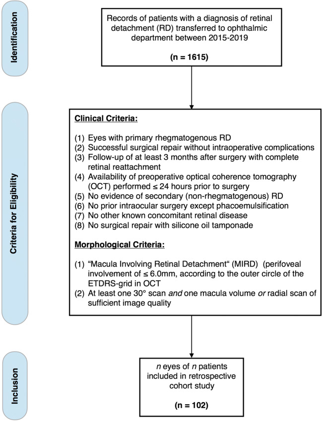
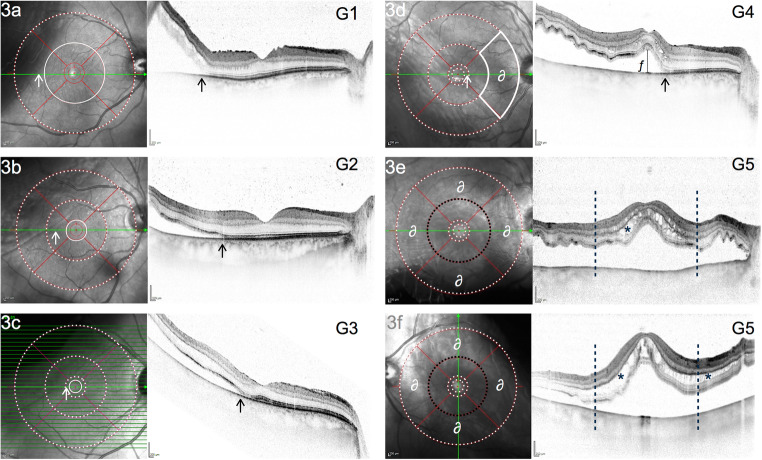
To introduce an ETDRS grid-based classification for macula involving retinal detachment (MIRD) with or without center (foveal) involvement and to identify biomarkers in preoperative optical coherence tomography (OCT) associated with a favorable postoperative functional outcome in eyes with center involving retinal detachment (CIRD). One hundred and two eyes of 102 consecutive patients (f/m: 35/67) with primary rhegmatogenous retinal detachment, preoperative evidence of MIRD (perifoveal involvement of ≤ 6.0 mm), and successful retinal surgery were included in this retrospective cohort study. Eyes were assigned to 5 grades of MIRD (G1-G5), based on the extent of detachment in the ETDRS grid. Eyes with a detached foveal status (CIRD) were assigned to G4 or G5. In CIRD, the following OCT biomarkers were quantified and correlated with mean BCVA (logMAR) at 3 months postsurgery, using univariate and multivariable regression models: grade of detachment, extent of intraretinal edema, height of foveal detachment, subretinal folds, and epiretinal membrane. Forty-one of 102 eyes (40.2%) presented with an attached foveal status, defined as either outer (G1: 11.8%) or inner (G2: 18.6%) macular involvement or fovea-threatening MIRD (G3: 9.8%). Sixty-one eyes (59.8%) showed CIRD (G4 or G5). Eyes with CIRD had significantly worse postoperative BCVA than eyes without foveal involvement (0.355 logMAR vs. 0.138 logMAR, p<0.001). If CIRD was limited to three outer ETDRS quadrants (G4), mean BCVA was better compared to CIRD involving all four ETDRS quadrants (G5) (0.254 logMAR vs. 0.522 logMAR, p<0.001). Reading ability (BCVA ≤ 0.4 logMAR) was restored in 97.6% of eyes with G1-G3 compared to 86.9% of eyes with G4 (p=0.072) and 52.4% of eyes with G5 (p<0.001). In multivariable regression analysis of eyes with CIRD, a lower grade of detachment (G4 vs. G5: p<0.05) and lower extent of cystoid edema (focal/none vs. wide: p<0.001) were both associated with better postoperative function. The functional outcome after MIRD may be worse in the presence of foveal involvement (CIRD), but a lower grade of detachment and the absence of intraretinal edema can predict a good recovery in spite of CIRD.
Keywords: Biomarker; CIRD; Center Involving Retinal; Detachment; Fovea-off; MIRD; Macula Involving Retinal Detachment; Macula-off; Macula-on; Morphology; Nomenclature; OCT; Optical coherence tomography; Retinal detachment; Rhegmatogenous.
© 2021. The Author(s).
Conflict of interest statement
J Klaas reports personal fees from Novartis, outside of the submitted work. N Feucht reports personal fees from Novartis, Allergan, Bayer, and Heidelberg Engineering, outside the submitted work. J Siedlecki received previous speaker fees and travel expenses from Novartis Pharma GmbH, Carl Zeiss Meditec AG, Oculentis OSD Medical GmbH, and Pharm-Allergan GmbH. Jakob Siedlecki received personal consultation fees from Novartis Pharma GmbH, Bayer AG, and Pharm-Allergan GmbH. Jakob Siedlecki received travel support from Oertli AG. M Maier reports speaker honoraria from Novartis, Allergan, Bayer, Heidelberg Engineering, and Zeiss, Clinical Trials (Bayer, Novartis, Roche), outside the submitted work. P Rechl, J Friedrich, and CP Lohmann have nothing to disclose.
Figures

References
-
- Klaas JE, Rechl P, Klein J, Feucht N, Lohmann CP, Maier M (2020) OCT Biomarker bei rhegmatogener Netzhautablösung mit Makulabeteiligung. Deutsche Ophthalmologische Gesellschaft. 10.1007/s00347-020-01298-w
MeSH terms
Substances
LinkOut - more resources
Full Text Sources
Other Literature Sources
Medical
