

Hair Loss Caused by Gain-of-Function Mutant TRPV3 Is Associated with Premature Differentiation of Follicular Keratinocytes
- PMID: 33675791
- PMCID: PMC8694045
- DOI: 10.1016/j.jid.2020.11.036
Hair Loss Caused by Gain-of-Function Mutant TRPV3 Is Associated with Premature Differentiation of Follicular Keratinocytes
Abstract
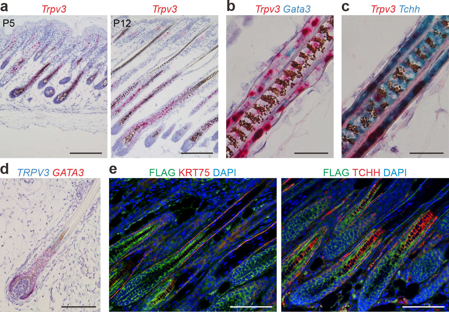
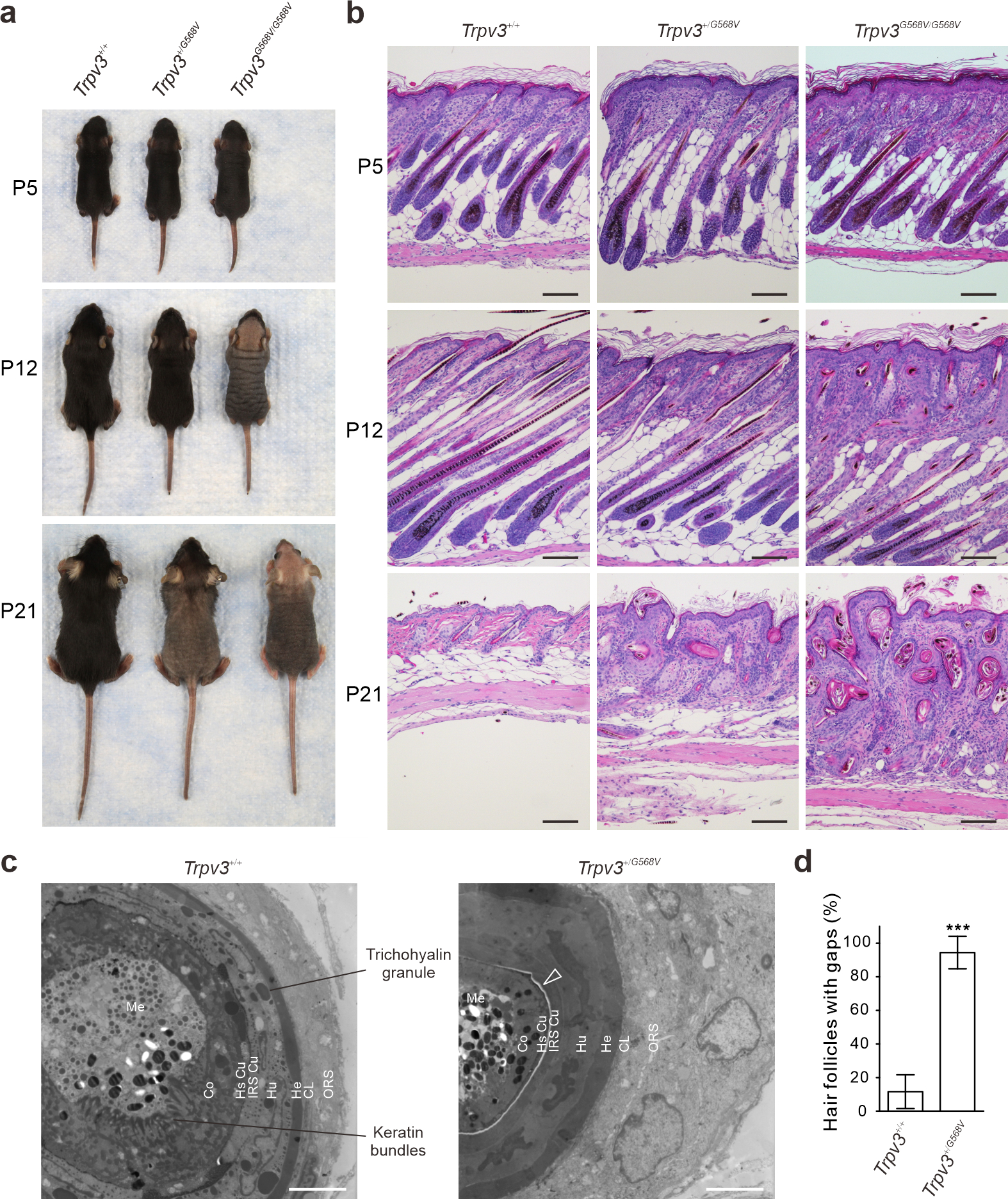
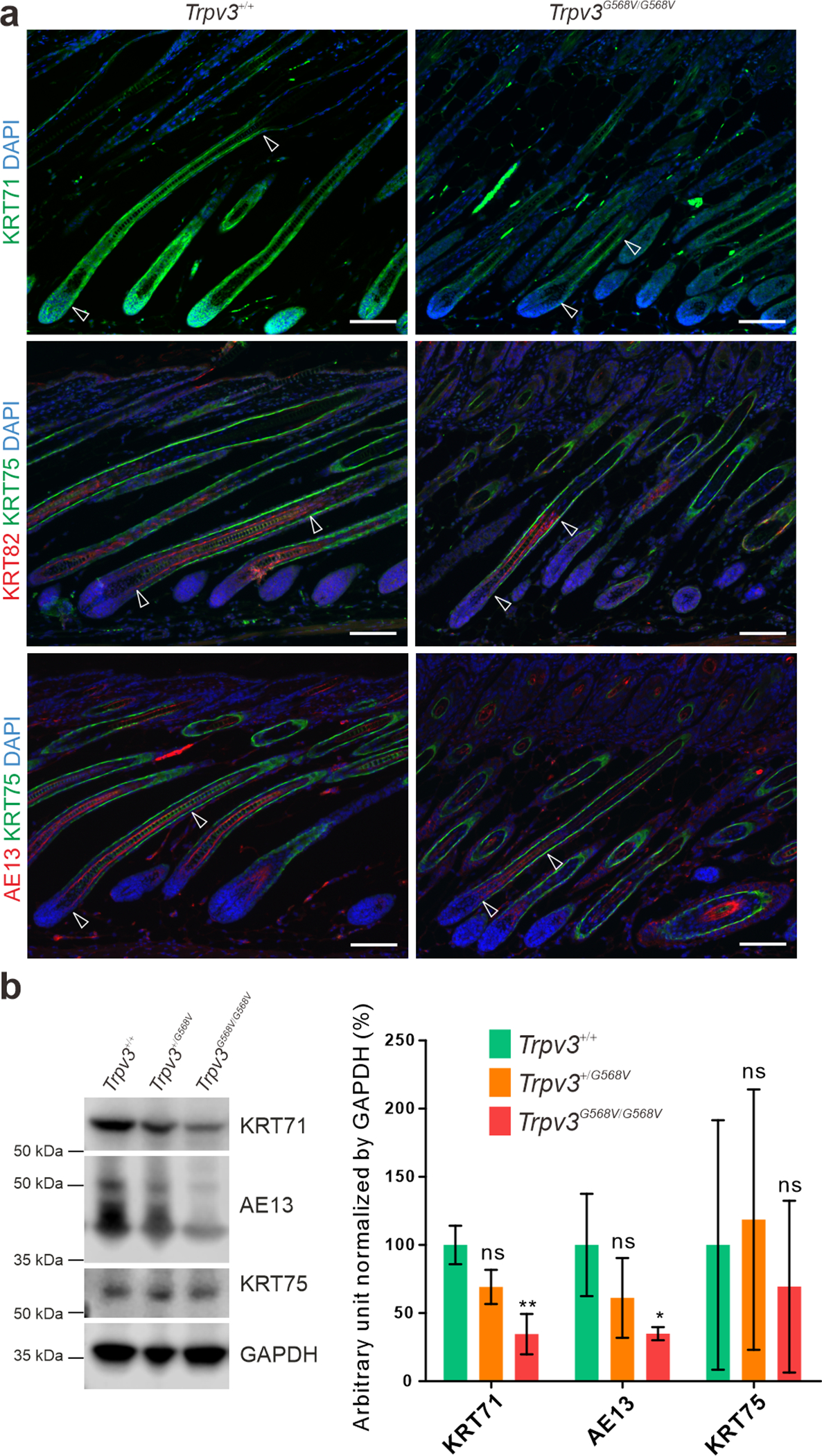
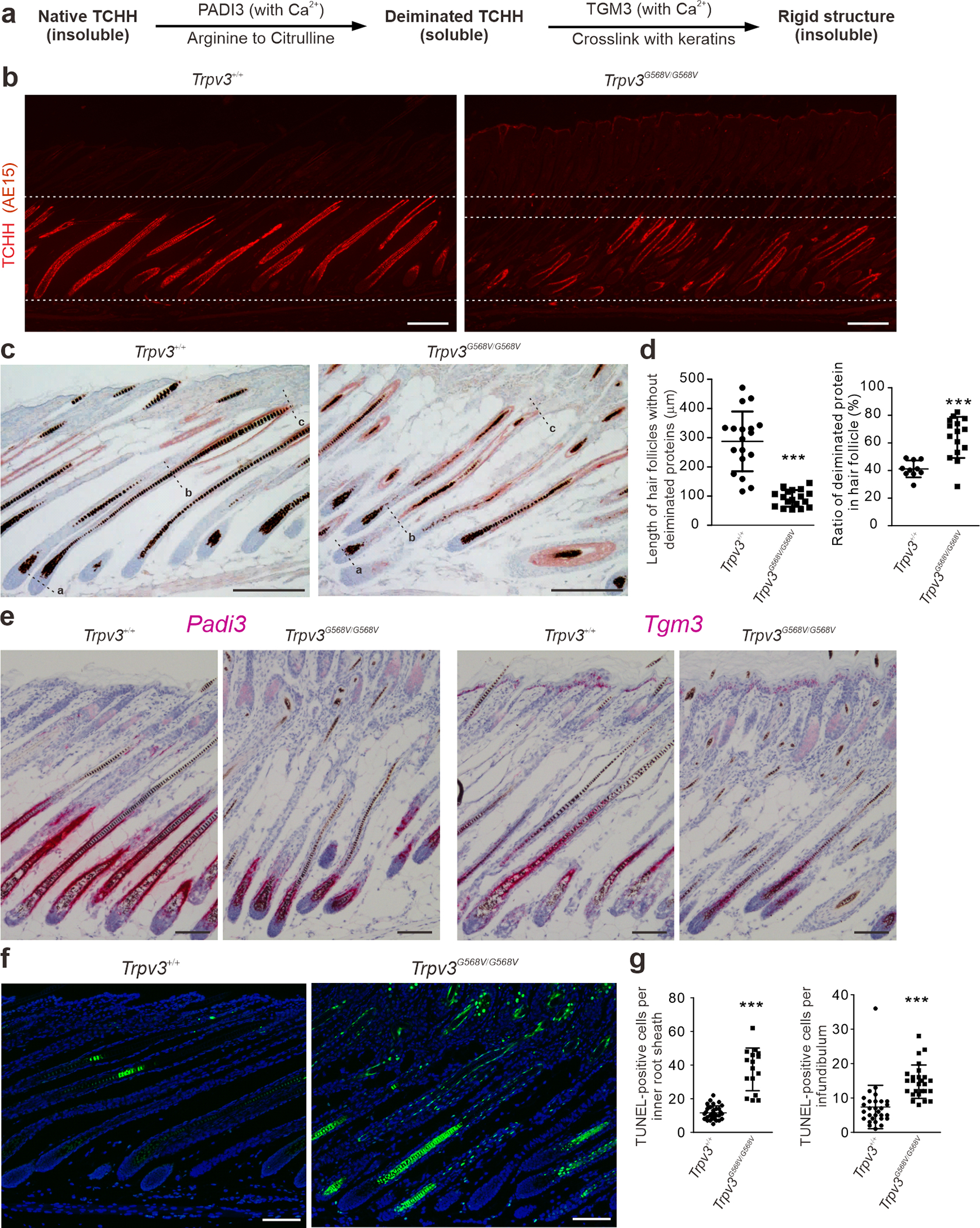
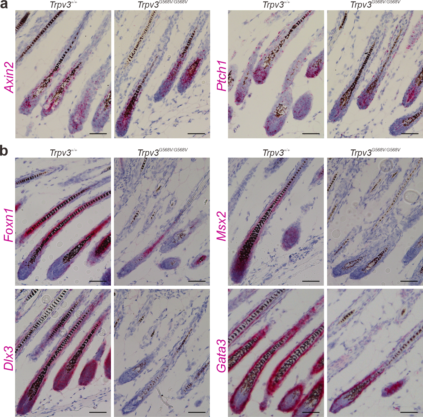
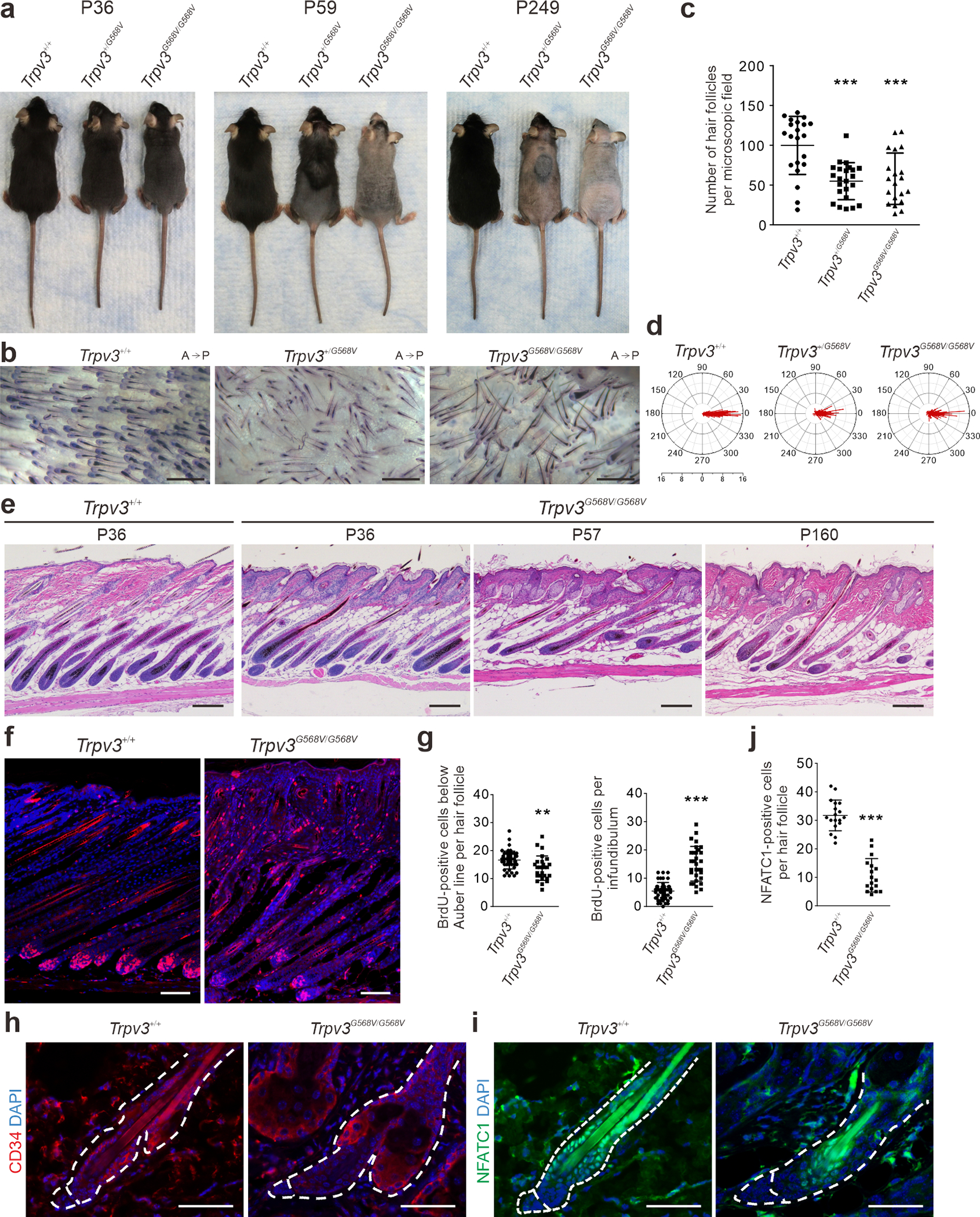
Gain-of-function mutations in the TRPV3 gene can cause Olmsted syndrome characterized by palmoplantar and periorificial keratoderma, itch, and hair loss. The mechanism underlying the hair loss remains unclear. In this study, we engineered an Olmsted syndrome mouse model by introducing the point mutation G568V to the corresponding Trpv3 locus in the mice. These mice developed fully penetrant hair loss. The hair loss was associated with premature differentiation of follicular keratinocytes characterized by precocious degeneration of trichohyalin and keratins, increased production of deiminated proteins, elevated apoptosis, and attenuation of transcription regulators (Foxn1, Msx2, Dlx3, and Gata3) known to regulate hair follicle differentiation. These abnormalities occurred in the medial‒proximal region of the inner root sheath and the hair shaft, where Trpv3 is highly expressed, and correlated with an impaired formation of the hair canal and the hair shaft. The mutant Trpv3 mice also exhibited increased proliferation in the outer root sheath, accelerated hair cycle, reduction of hair follicle stem cells, and miniaturization of regenerated hair follicles. Findings from this study suggest that precocious maturation of postmitotic follicular keratinocytes drives hair loss in patients with Olmsted syndrome.
Copyright © 2021 The Authors. Published by Elsevier Inc. All rights reserved.
Conflict of interest statement
CONFLICT OF INTEREST
The authors state no conflict of interest.
Figures
Similar articles
-
Exome sequencing reveals mutations in TRPV3 as a cause of Olmsted syndrome.Am J Hum Genet. 2012 Mar 9;90(3):558-64. doi: 10.1016/j.ajhg.2012.02.006. Am J Hum Genet. 2012. PMID: 22405088 Free PMC article.
-
Pharmacological Activation of Thermo-Transient Receptor Potential Vanilloid 3 Channels Inhibits Hair Growth by Inducing Cell Death of Hair Follicle Outer Root Sheath.J Pharmacol Exp Ther. 2019 Aug;370(2):299-307. doi: 10.1124/jpet.119.258087. Epub 2019 May 31. J Pharmacol Exp Ther. 2019. PMID: 31152005
-
Olmsted Syndrome Caused by a Heterozygous p.Gly568Val Missense Mutation in TRPV3 Gene.Yonsei Med J. 2018 Mar;59(2):341-344. doi: 10.3349/ymj.2018.59.2.341. Yonsei Med J. 2018. PMID: 29436206 Free PMC article.
-
Novel Insights into the Role of Keratinocytes-Expressed TRPV3 in the Skin.Biomolecules. 2023 Mar 10;13(3):513. doi: 10.3390/biom13030513. Biomolecules. 2023. PMID: 36979447 Free PMC article. Review.
-
TRPV3 Mutation-Associated Olmsted Syndrome in a Hispanic Patient: Response to Erlotinib and Review of Literature.Pediatr Dermatol. 2025 Jul-Aug;42(4):835-840. doi: 10.1111/pde.15892. Epub 2025 Feb 5. Pediatr Dermatol. 2025. PMID: 39910762 Review.
Cited by
-
Th2 Modulation of Transient Receptor Potential Channels: An Unmet Therapeutic Intervention for Atopic Dermatitis.Front Immunol. 2021 Jun 30;12:696784. doi: 10.3389/fimmu.2021.696784. eCollection 2021. Front Immunol. 2021. PMID: 34276687 Free PMC article. Review.
-
5'tiRNA-Glu-TTC targets TRPV3 and activates the PI3K/AKT signaling pathway to modulate skin photoaging.Noncoding RNA Res. 2025 Jul 14;15:29-43. doi: 10.1016/j.ncrna.2025.07.004. eCollection 2025 Dec. Noncoding RNA Res. 2025. PMID: 40741358 Free PMC article.
-
Integration Analysis of Transcriptome Sequencing and Whole-Genome Resequencing Reveal Wool Quality-Associated Key Genes in Zhexi Angora Rabbits.Vet Sci. 2024 Dec 13;11(12):651. doi: 10.3390/vetsci11120651. Vet Sci. 2024. PMID: 39728991 Free PMC article.
-
Advances in the Study for Modulators of Transient Receptor Potential Vanilloid (TRPV) Channel Family.Curr Top Med Chem. 2025;25(12):1403-1450. doi: 10.2174/0115680266294569241115053420. Curr Top Med Chem. 2025. PMID: 39757673 Review.
-
Latest Insights into the In Vivo Studies in Murine Regarding the Role of TRP Channels in Wound Healing-A Review.Int J Mol Sci. 2024 Jun 19;25(12):6753. doi: 10.3390/ijms25126753. Int J Mol Sci. 2024. PMID: 38928459 Free PMC article. Review.
References
-
- Alibardi L Fine structure and immunocytochemistry of monotreme hairs, with emphasis on the inner root sheath and trichohyalin-based cornification during hair evolution. J Morphol 2004;261(3):345–63. - PubMed
-
- Asakawa M, Yoshioka T, Matsutani T, Hikita I, Suzuki M, Oshima I, et al. Association of a mutation in TRPV3 with defective hair growth in rodents. J Invest Dermatol 2006;126(12):2664–72. - PubMed
-
- Borbiro I, Lisztes E, Toth BI, Czifra G, Olah A, Szollosi AG, et al. Activation of transient receptor potential vanilloid-3 inhibits human hair growth. J Invest Dermatol 2011;131(8):1605–14. - PubMed
-
- Candi E, Schmidt R, Melino G. The cornified envelope: a model of cell death in the skin. Nat Rev Mol Cell Biol 2005;6(4):328–40. - PubMed
Publication types
MeSH terms
Substances
Grants and funding
LinkOut - more resources
Full Text Sources
Other Literature Sources
Molecular Biology Databases
Research Materials
