. 2021 Mar 6;13(1):57.
doi: 10.1186/s13195-021-00791-x.

Human neural stem cell-derived extracellular vesicles mitigate hallmarks of Alzheimer's disease

Affiliations

Human neural stem cell-derived extracellular vesicles mitigate hallmarks of Alzheimer's disease

Lauren A Apodaca et al. Alzheimers Res Ther. .

Abstract

Background: Regenerative therapies to mitigate Alzheimer's disease (AD) neuropathology have shown very limited success. In the recent era, extracellular vesicles (EVs) derived from multipotent and pluripotent stem cells have shown considerable promise for the treatment of dementia and many neurodegenerative conditions.

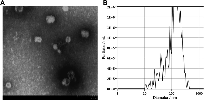
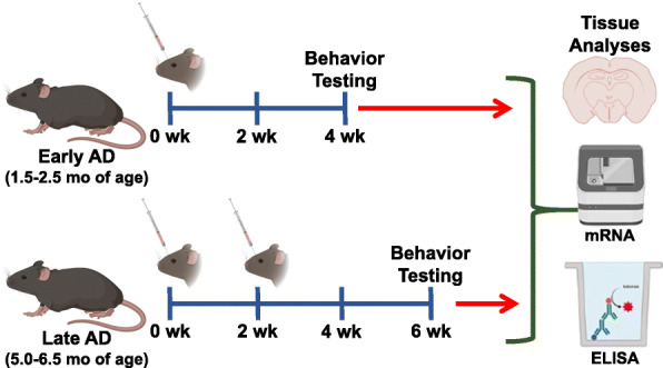
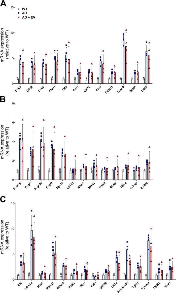
Methods: Using the 5xFAD accelerated transgenic mouse model of AD, we now show the regenerative potential of human neural stem cell (hNSC)-derived EVs on the neurocognitive and neuropathologic hallmarks in the AD brain. Two- or 6-month-old 5xFAD mice received single or two intra-venous (retro-orbital vein, RO) injections of hNSC-derived EVs, respectively.

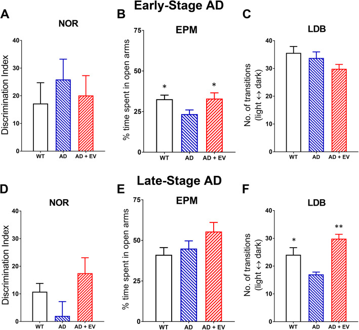
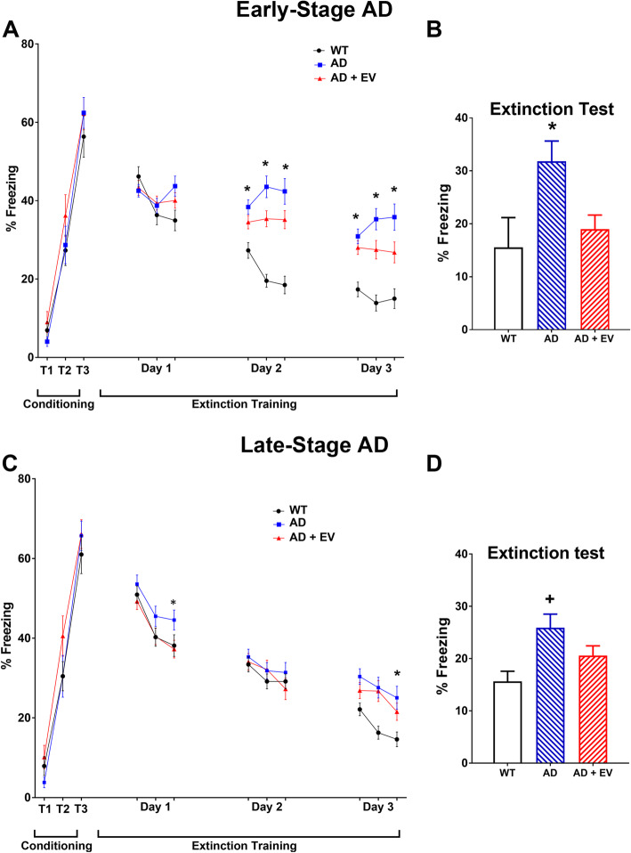
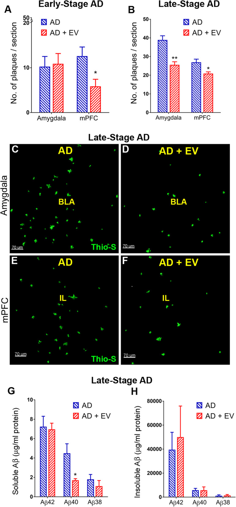
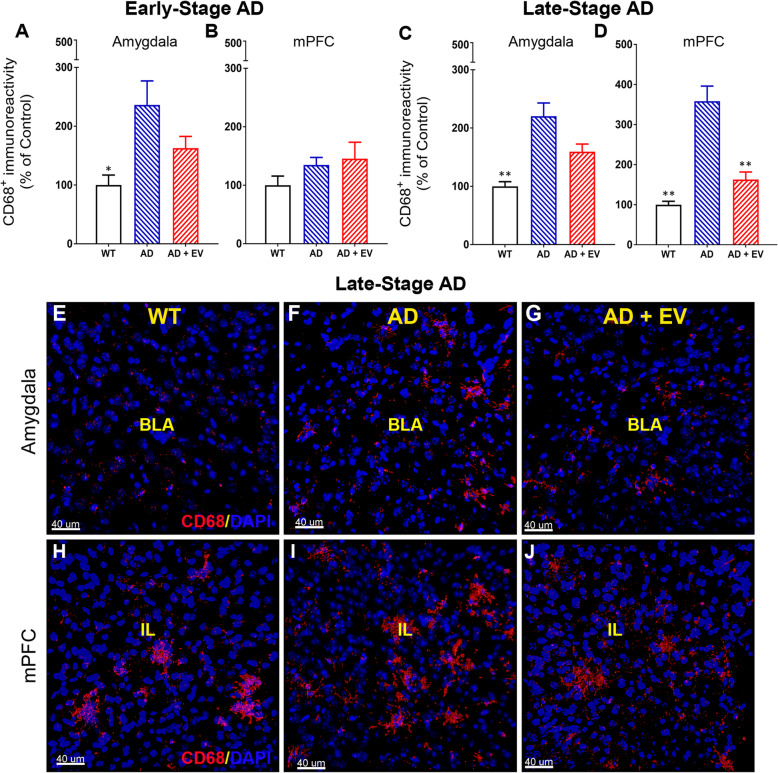
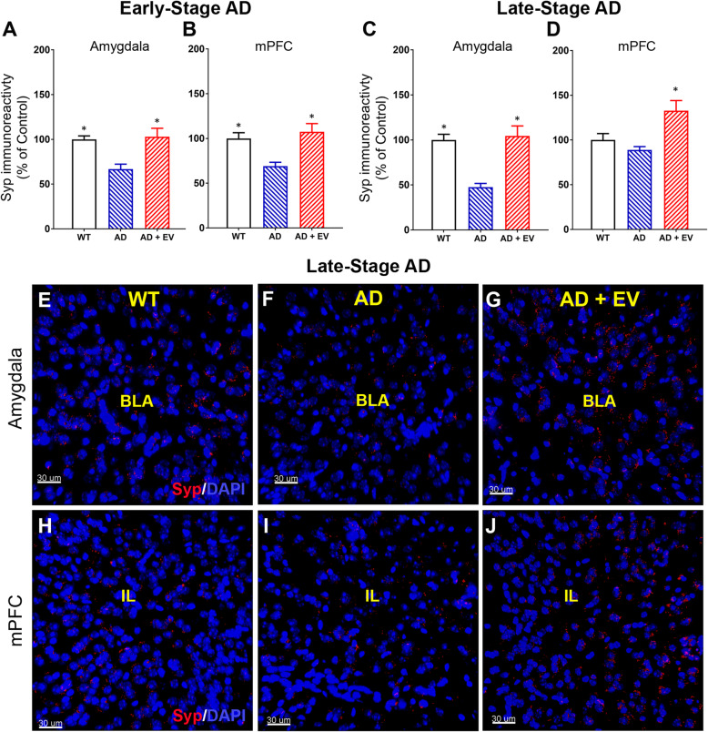
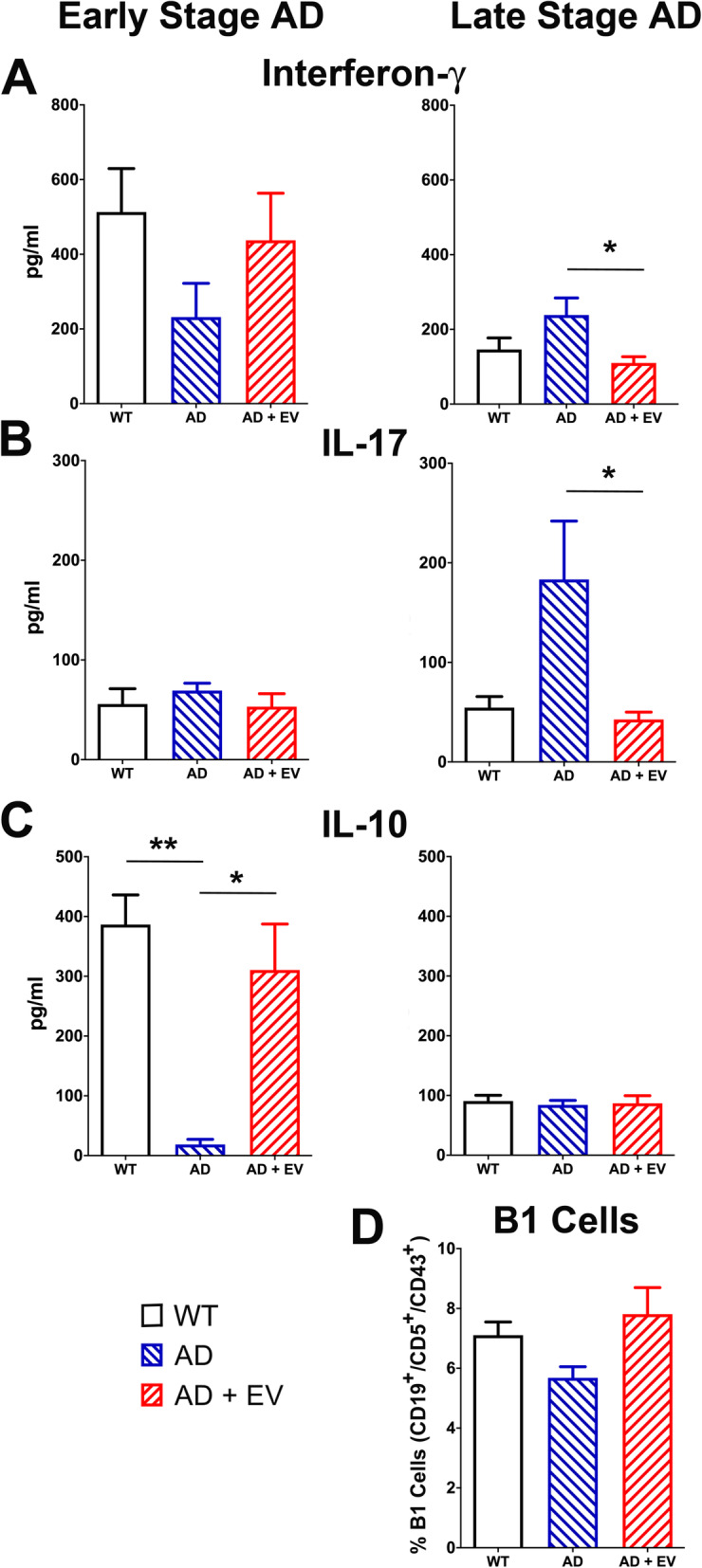
Results: RO treatment using hNSC-derived EVs restored fear extinction memory consolidation and reduced anxiety-related behaviors 4-6 weeks post-injection. EV treatment also significantly reduced dense core amyloid-beta plaque accumulation and microglial activation in both age groups. These results correlated with partial restoration of homeostatic levels of circulating pro-inflammatory cytokines in the AD mice. Importantly, EV treatment protected against synaptic loss in the AD brain that paralleled improved cognition. MiRNA analysis of the EV cargo revealed promising candidates targeting neuroinflammation and synaptic function.

Conclusions: Collectively, these data demonstrate the neuroprotective effects of systemic administration of stem cell-derived EVs for remediation of behavioral and molecular AD neuropathologies.

Keywords: Alzheimer’s disease; Extracellular vesicle; Inflammatory response; Neural stem cell.


Conflict of interest statement

The authors declare no competing interests.

Figures

Fig. 1
Fig. 1
Characterization of extracellular vesicles. a Representative electron microscopy image depicts typical EV morphology and size. b Representative graph from ZetaView analysis showing the average size distribution of the EVs
Fig. 2
Fig. 2
Experimental design. Early AD male mice were treated with a single dose of human neural stem cell-derived extracellular vesicles (EVs) via retro-orbital vein (RO) injection at ~ 1.5–2.5 months of age and behaviorally tested 1 month later. Late-stage AD male mice received 2 RO injections of EVs at 2-week intervals and began behavior testing 1 month after the second injection
Fig. 3
Fig. 3
ac Early-stage AD behavioral testing indicated no impairments on novel object recognition (NOR) or light-dark box (LDB) tests, but AD mice showed increased anxiety-like behavior on the elevated plus maze (EPM) test that was mitigated by EV treatment. df Late-stage AD mice exhibited no significant impairments on NOR or EPM tests, but increased anxiety-like behavior on the LDB test that was mitigated by EV treatment as shown by an increased number of transitions between the light-dark compartments. Data are presented as mean ± SEM where N = 14–16 mice/group. P values are derived from ANOVA and Bonferroni’s multiple comparisons test. *P < 0.05, **P < 0.01 as compared to AD
Fig. 4
Fig. 4
AD mice exhibited impairments in fear extinction memory and enhanced memory recall. All mice showed elevated freezing following a series of 3 tone-shock pairings (0.6 mA, T1–T3). Subsequently, fear extinction training was administered every 24 h (15 tones) for 3 days. These data are presented as the average of 5 tones (a, c). All a early and c late study mice exhibit a gradual decrease in freezing behavior (days 1–3); however, AD mice spent significantly more time freezing during the extinction training compared to controls. Twenty-four hours after extinction training, b early-stage AD mice exhibited enhanced fear recall (elevated freezing, P < 0.05), while AD mice receiving EV treatment exhibited successful extinction equivalent to that of WT control mice (reduced freezing). d Late-stage AD mice exhibited enhanced fear recall as compared to WT control mice. Data are presented as mean ± SEM where N = 14–16 mice/group. P values are derived from ANOVA and Bonferroni’s multiple comparisons test. *P < 0.05 as compared to WT and AD+EV, +P < 0.05 as compared to WT.
Fig. 5
Fig. 5
Dense core Aβ plaque number was reduced in AD mice that received EV treatment. a Early-stage AD mice exhibited increased numbers of Aβ plaques in the amygdala that were reduced by EV treatment and b late-stage AD mice exhibited increased numbers of Aβ plaques in both the amygdala and the medial prefrontal cortex (mPFC) that were reduced by EV treatment. Representative images of Thio-S staining for late-stage AD and AD+EV mice qualitatively demonstrate late AD neuropathology as shown by the accumulation of Aβ plaques in the c amygdala and e mPFC that were reduced in both the d amygdala and f mPFC regions of the AD brain by EV treatment (basal lateral amygdala, BLA; infralimbic cortex, IL; ABP, green). g Aβ multiplex ELISA indicated the elevation of soluble Aβ40 in brain lysates that was significantly reduced by EV treatment, h while no changes were observed between AD and AD+EV mice in levels of insoluble Aβ. Data are presented as mean ± SEM (Thio-S, N = 4 mice/group; ELISA, N = 7 mice/group). P values derived from unpaired Student’s t tests. *P < 0.05, **P < 0.01 as compared to AD. Scale bar = 70 μm
Fig. 6
Fig. 6
Microglial activation was reduced in AD mice that received EV treatment. Early-stage AD mice exhibited significantly increased numbers of CD68+ microglia in the a amygdala that were not significantly altered by EV treatment, and b no effect of disease or treatment was observed in the medial prefrontal cortex (mPFC). Late-stage AD mice showed significant increases in CD68+ immunoreactivity in the c amygdala and d mPFC that were ameliorated by EV treatment. Representative images of CD68 staining for late-stage mice qualitatively demonstrate these relative changes in eg the amygdala of wild type, AD, and AD mice treated with EVs (WT, AD, AD+EV, respectively) and hj the mPFC similarly (basal lateral amygdala, BLA; infralimbic cortex, IL; red, CD68; blue, DAPI nuclear counterstain). Data are presented as mean ± SEM (N = 4 mice/group). P values are derived from ANOVA and Bonferroni’s multiple comparisons test. *P < 0.05, **P < 0.01  as compared to AD. Scale bar = 40 μm
Fig. 7
Fig. 7
Levels of the pre-synaptic protein synaptophysin were reduced in the brains of AD mice. Evaluation of synaptophysin (Syp) immunoreactivity revealed significant decreases in the a amygdala and the b medial prefrontal cortex (mPFC) of early-stage AD mice that were ameliorated by EV treatment. Late-stage AD mice showed significant reductions in Syp in the c amygdala that were ameliorated by EV treatment. d AD-related changes in Syp were not observed in the mPFC. Representative images of Syp staining from late-stage mice qualitatively demonstrate these relative changes in eg the amygdala of wild type, AD, and AD mice treated with EVs (WT, AD, AD+EV, respectively) and hj the mPFC similarly (basal lateral amygdala, BLA; infralimbic cortex, IL; red, Syp; blue, DAPI nuclear counterstain). Data are presented as mean ± SEM (N = 4 mice/group). P values are derived from ANOVA and Bonferroni’s multiple comparisons test. *P < 0.05  as compared to AD. Scale bar = 40 μm
Fig. 8
Fig. 8
Elevated levels of inflammatory cytokines in AD mice were reduced by EV treatment. Levels of cytokines secreted by PMA- and ionomycin-stimulated spleen cells were measured in WT, AD, and AD+EV mice. a, b While unaffected in early-stage AD mice, interferon-γ and IL-17 pro-inflammatory cytokines were significantly elevated in late-stage AD mice and significantly reduced by EV treatment. c Alternatively, reduced levels of the anti-inflammatory cytokine IL-10 in early-stage AD mice were restored to nearly control levels in the AD mice that received EV treatment. No changes among groups were observed for IL-10 in the late-stage animals. d Spleen cells were also stained for B1 cells (CD19+, CD5+, CD43+) and analyzed by flow cytometry. While not statistically significant, a trend for a reduced percentage of B1 cells was observed for AD mice and improved by EV treatment. Data are presented as mean ± SEM (N = 7 mice/group). P values are derived from ANOVA and Bonferroni’s multiple comparisons test. *P < 0.05, **P < 0.01
Fig. 9
Fig. 9
Gene expression in the hippocampus of late-stage AD mice. Analysis of a microglial, b pro-inflammation, c immune response, and other mRNA levels demonstrated increased gene expression in the hippocampus of late-stage AD mice as compared to WT controls. Those elevated mRNA levels were reduced in some, but not all AD+EV-treated mice. Data are presented as mean ± SEM (N = 4 mice/group)

References

    1. LaFerla FM, Green KN. Animal models of Alzheimer disease. Cold Spring Harb Perspect Med. 2012;2(11):a006320. doi: 10.1101/cshperspect.a006320. - DOI - PMC - PubMed
    1. Alam Q, Alam MZ, Mushtaq G, Damanhouri GA, Rasool M, Kamal MA, Haque A. Inflammatory process in Alzheimer’s and Parkinson’s diseases: central role of cytokines. Curr Pharm Des. 2016;22:541–548. doi: 10.2174/1381612822666151125000300. - DOI - PubMed
    1. Baulch JE, Acharya MM, Agrawal S, Apodaca LA, Monteiro C, Agrawal A. Immune and inflammatory determinants underlying Alzheimer’s disease pathology. J NeuroImmune Pharmacol. 2020;15(4):852–862. doi: 10.1007/s11481-020-09908-9. - DOI - PMC - PubMed
    1. Spangenberg EE, Lee RJ, Najafi AR, Rice RA, Elmore MR, Blurton-Jones M, West BL, Green KN. Eliminating microglia in Alzheimer’s mice prevents neuronal loss without modulating amyloid-beta pathology. Brain. 2016;139:1265–1281. doi: 10.1093/brain/aww016. - DOI - PMC - PubMed
    1. Parihar VK, Pasha J, Tran KK, Craver BM, Acharya MM, Limoli CL. Persistent changes in neuronal structure and synaptic plasticity caused by proton irradiation. Brain Struct Funct. 2014;220(2):1161–71. doi: 10.1007/s00429-014-0709-9. - DOI - PMC - PubMed

Publication types