. 2021 Mar 6;11(1):50.
doi: 10.1186/s13578-021-00563-y.

ApoE4 (Δ272-299) induces mitochondrial-associated membrane formation and mitochondrial impairment by enhancing GRP75-modulated mitochondrial calcium overload in neuron

Affiliations

ApoE4 (Δ272-299) induces mitochondrial-associated membrane formation and mitochondrial impairment by enhancing GRP75-modulated mitochondrial calcium overload in neuron

Tao Liang et al. Cell Biosci. .

Abstract

Background: Apolipoprotein E4 (apoE4) is a major genetic risk factor of Alzheimer's disease. Its C-terminal-truncated apoE4 (Δ272-299) has neurotoxicity by affecting mitochondrial respiratory function. However, the molecular mechanism(s) underlying the action of apoE4 (Δ272-299) in mitochondrial function remain poorly understood.

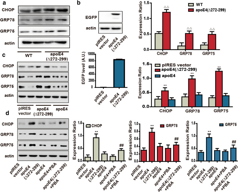
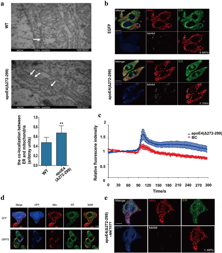
Methods: The impact of neuronal apoE4 (Δ272-299) expression on ER stress, mitochondrial-associated membrane (MAM) formation, GRP75, calcium transport and mitochondrial impairment was determined in vivo and in vitro. Furthermore, the importance of ER stress or GRP75 activity in the apoE4 (Δ272-299)-promoted mitochondrial dysfunction in neuron was investigated.

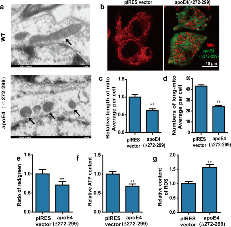
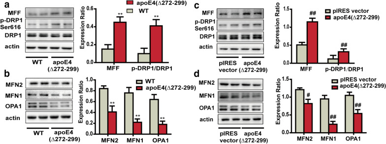
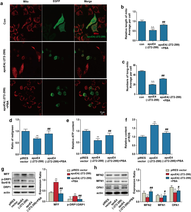
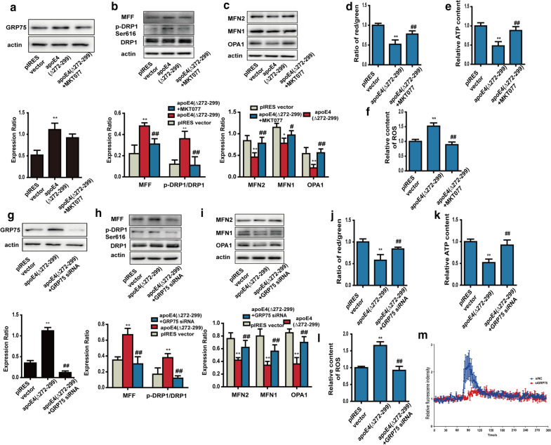
Results: Neuronal apoE4 (Δ272-299) expression induced mitochondrial impairment by inducing ER stress and mitochondrial-associated membrane (MAM) formation in vivo and in vitro. Furthermore, apoE4 (Δ272-299) expression promoted GRP75 expression, mitochondrial dysfunction and calcium transport into the mitochondria in neuron, which were significantly mitigated by treatment with PBA (an inhibitor of ER stress), MKT077 (a specific GRP75 inhibitor) or GRP75 silencing.

Conclusions: ApoE4 (Δ272-299) significantly impaired neuron mitochondrial function by triggering ER stress, up-regulating GRP75 expression to increase MAM formation, and mitochondrial calcium overload. Our findings may provide new insights into the neurotoxicity of apoE4 (Δ272-299) against mitochondrial function and uncover new therapeutic targets for the intervention of Alzheimer's disease.

Keywords: Alzheimer’s disease; Apolipoprotein E4; ER stress; Mitochondrial Ca2+ overload; Mitochondria‐associated ER membrane; apoE4 (Δ272–299).


Conflict of interest statement

The authors declare no competing interests.

Figures

Fig. 1
Fig. 1
ApoE4 (Δ272–299) triggers ER stress in vivo and in vitro. a Western blot analysis the relative levels of CHOP, GRP78 and GRP75 expression in the hippocampus of apoE4 (Δ272–299) transgenic and the age- and sex-matched wild-type mice. b Transfection with pEGFP-apoE4 (Δ272–299) induces EGFP expression in N2a cells. c Western blot analysis of the relative levels of CHOP, GRP78 and GRP75 expression in the different groups of N2a cells. d Treatment with PBA mitigated the apoE4 (Δ272–299)-up-regulated CHOP, GRP78 and GRP75 expression in N2a cells (ΔΔP < 0.01 versus the wild-type mice; **P < 0.01 versus the N2a cells transfected with pIRES vector; ##P < 0.01 versus the apoE4 (Δ272–299)-expressed cells). Data are representative images or expressed as the mean ± SEM of each group (n = 3) from three separate experiments. The significant differences were determined by ANOVA and post hoc Fisher’s least significant difference
Fig. 2
Fig. 2
ApoE4 (Δ272–299) induces mitochondrial morphology change and dysfunction. a TEM analysis of the morphological changes in mitochondria in the hippocampus of wild-type mice and apoE4 (Δ272–299) transgenic mice. b N2a cells were tragnsiently transfected with pIRES or pIRES-apoE4 (Δ272–299) for 24 h, and their mitochondria were stained with mitotracker. The morphological changes in mitochondria were examined under a laser confocal microscope. The scale bar is 10 µm. c The length of mitochondria was measured using the ImageJ, n = 20–30 mitochondria per group. d The areas of mitochondria was also measured using the ImageJ, n = 20–30 cells. e The MMP was detected by the JC-1. f The levels of ATP were analyzed by chemiluminescence. g The levels of mitochondrial ROS were analyzed by DCFH-DA. Data are representative images or expressed as the mean ± SEM of each group (n = 3) from three separate experiments. **P < 0.01 versus the control N2a cells transfected with pIRES vector, The significant differences were determined by ANOVA and post hoc Fisher’s least significant difference
Fig. 3
Fig. 3
ApoE4 (Δ272–299) induces the imbalance of mitochondrial fusion and fission in vivo and in vitro. a, b Western blot analysis of the relative levels of MFF, MFN2, MFN1 and OPA1 expression and DRP1 phosphorylation in the hippocampus of apoE4 (Δ272–299) transgenic and wild-type mice. c, d Western blot analysis of the relative levels of MFF, MFN2, MFN1 and OPA1 expression and DRP1 phosphorylation in the apoE4 (Δ272–299) expressed N2a and control cells. Data are representative images or expressed as the mean ± SEM of each group (n = 3) from three separate experiments. **P < 0.01 versus the wild-type mice; ##P < 0.01, #P < 0.05 versus the control N2a cells transfected with the pIRES vector. The significant differences were determined by ANOVA and post hoc Fisher’s least significant difference
Fig. 4
Fig. 4
PBA treatment mitigates the apoE4 (272–299)-induced mitochondrial impairment in N2a cells. N2a cells were transiently transfected with apoE4 (Δ272–299), and were treated with, or without, 1 mM PBA for another 18 h. a The mitochondria of N2a cells were stained with mitotracker, and were examined by a laser confocal microscope. The scale bar is 10 µm. b The lengths of mitochondria were measured using the ImageJ, n = 20–30 mitochondria per group. c The areas of mitochondria were measured using the ImageJ, n = 20–30 cells per group. d The MMP was analyzed by the JC-1. e The levels of ATP were analyzed by chemiluminescence. f The levels of ROS were analyzed by DCFH-DA. g, h Western blot analysis of the relative levels of MFF, MFN2, MFN1 and OPA1 expression and DRP1 phosphorylation in the difference groups of N2a cells. ΔΔP < 0.01 versus the control N2a cells; **P < 0.01,*P < 0.05, versus the vector-transfected N2a cells; ##P < 0.01, #P < 0.05, versus the apoE4 (Δ272–299) expressed N2a cells. Data are representative images or expressed as the mean ± SEM of each group (n = 3) from three separate experiments. The significant differences were determined by ANOVA and post hoc Fisher’s least significant difference
Fig. 5
Fig. 5
ApoE4 (Δ272–299) promotes ER-mitochondria interaction and mitochondrial Ca2+ overload. a TEM analysis of the MAM in the hippocampus of wild-type and apoE4 (Δ272–299) transgenic mice. b Fluorescent microscopy analysis of MAM in apoE4 (Δ272–299)-expressed N2a cells. The scale bar is 10 µm. c Longitudinal analysis of Ca2+ levels in mitochondria of N2a cells following inducing apoE4 (Δ272–299) expression. d Induction of GRP75 over-expression increased the contact areas of MAM in N2a cells. The scale bar is 10 µm. e Treatment with MKT077 mitigated the MAM changes induced by apoE4 (Δ272–299) in N2a cells. The scale bar is 10 µm. Data are representative images or expressed as the mean ± SEM of each group (n = 3) from three separate experiments. **P < 0.01, versus the wild-type mice. The significant differences were determined by ANOVA and post hoc Fisher’s least significant difference
Fig. 6
Fig. 6
Inhibition of GRP75 alleviates the apoE4 (Δ272–299)-induced mitochondrial impairment in N2a cells. N2a cells were transiently transfected with the plasmid pIRES or apoE4 (Δ272–299), and treated with, or without, 1 µM GRP75 inhibitor MKT077 for 18 h. Furthermore, the apoE4 (Δ272–299)-transfected N2a cells were transfected with 50 nM control or GRP75-specific siRNA for 18 h. a, g Western blot analysis of the relative levels of GRP75 expression in N2a cells. b, c, h, i Western blot analysis of the relative levels of MFF, MFN2, MFN1 and OPA1 expression and DRP1 phosphorylation in N2a cells. d, j The MMP was analyzed by the JC-1. e, k The levels of ATP were analyzed by chemiluminescence. f, l The levels of ROS were analyzed by DCFH-DA. m Longitudinal analysis of Ca2+ levels in mitochondria of N2a cells. Data are representative images or expressed as the mean ± SEM of each group (n = 3) from three separate experiments. **P < 0.01, *P < 0.05 versus the control N2a cells; ##P < 0.01, #P < 0.05 versus the apoE4 (Δ272–299)-expressed N2a cells. The significant differences were determined by ANOVA and post hoc Fisher’s least significant difference

Similar articles

Cited by

References

    1. Kim JH, Oho AUID- Genetics of Alzheimer’s Disease. Dement Neurocogn Disord. 2018;17:131–6. doi: 10.12779/dnd.2018.17.4.131. - DOI - PMC - PubMed
    1. Bellenguez C, Grenier-Boley B, Lambert JC. Genetics of Alzheimer’s disease: where we are, and where we are going. Curr Opin Neurobiol. 2020;61:40–8. doi: 10.1016/j.conb.2019.11.024. - DOI - PubMed
    1. Corder EH, Saunders AM, Strittmatter WJ, Schmechel DE, Gaskell PC, Small GW, Roses AD, Haines JL, Pericak-Vance MA. Gene dose of apolipoprotein E type 4 allele and the risk of Alzheimer’s disease in late onset families. Science. 1993;261:921–3. doi: 10.1126/science.8346443. - DOI - PubMed
    1. Tesseur I, Van Dorpe J, Spittaels K, Van den Haute C, Moechars D, Van Leuven F. Expression of human apolipoprotein E4 in neurons causes hyperphosphorylation of protein tau in the brains of transgenic mice. Am J Pathol. 2000;156:951–64. doi: 10.1016/S0002-9440(10)64963-2. - DOI - PMC - PubMed
    1. Tesseur I, Van Dorpe J, Bruynseels K, Bronfman F, Sciot R, Van Lommel A, Van Leuven F. Prominent axonopathy and disruption of axonal transport in transgenic mice expressing human apolipoprotein E4 in neurons of brain and spinal cord. Am J Pathol. 2000;157:1495–510. doi: 10.1016/S0002-9440(10)64788-8. - DOI - PMC - PubMed

LinkOut - more resources