

doi: 10.1016/j.chom.2021.02.017.
Epub 2021 Feb 24.
One year of SARS-CoV-2 evolution
Affiliations
- PMID: 33676588
- PMCID: PMC7903908
- DOI: 10.1016/j.chom.2021.02.017
Item in Clipboard
One year of SARS-CoV-2 evolution
Cell Host Microbe.
.
Abstract
Since the outbreak of SARS-CoV-2, the etiologic agent of the COVID-19 pandemic, the viral genome has acquired numerous mutations with the potential to increase transmission. One year after its emergence, we now further analyze emergent SARS-CoV-2 genome sequences in an effort to understand the evolution of this virus.
Copyright © 2021. Published by Elsevier Inc.
Figures
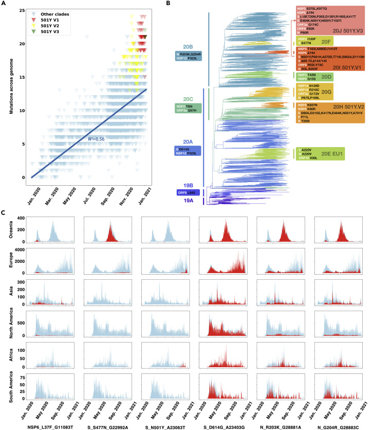
Phylogenetic pathway and spatiotemporal distribution of accumulated mutations in the SARS-CoV-2 genomes (A) The accumulated mutations in SARS-CoV-2 strains compared with early reference strain EPI_ISL_402125 since January of 2021. The 501Y.V1, 501Y.V2, and 501Y.V3 sub-clades were colored as red, yellow, and green, respectively. The linear regression line was shown and labeled. (B) Phylogenetic tree with fixed amino acid mutations for representative SARS-CoV-2 strains selected by Nextstrain until January 11th, 2021. Fixed mutations detected in each cluster were displayed in boxes. Viral strains were divided into hierarchical clusters as those in Nextstrain, including four big clusters (19A, 19B, 20A, and 20B) and eight small clusters (20C, 20D, 20E, 20F, 20G, 20H, 20I, and 20J). (C) The spatiotemporal distribution of SARS-CoV-2 genomes collected in GISAID with the six representative mutations during January 2020 to January 2021. In the histogram for each continent/mutation pairing, the x axis represents the collection date of the sequenced viruses and the y axis represents the number of sequences with the indicated mutation. The number of mutated sequences is shown in red, with the total number of all strains shown in light blue for comparison.
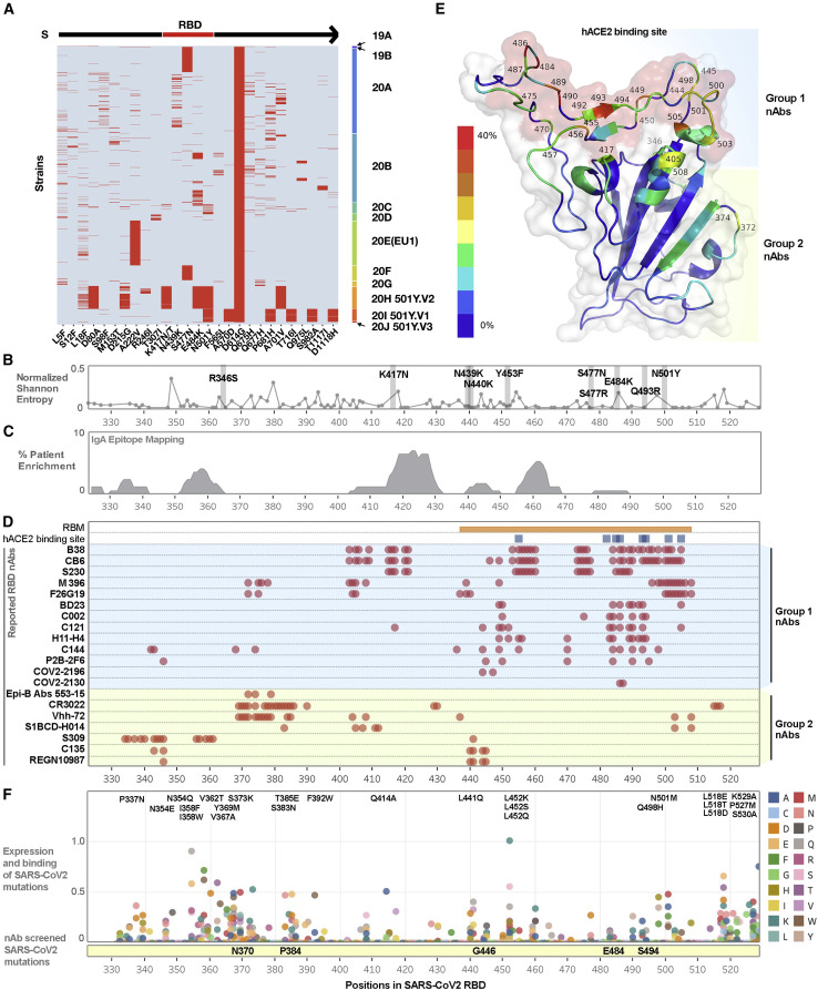
Systematic analysis of SARS-CoV-2 RBD mutations and neutralizing antibodies (A) Recurrent (appeared more than 20 times or >0.52% in the representative sequences) amino acid mutations in the S protein of SARS-CoV-2. To more clearly display the varied mutations in each strain, we show only those viral strains with the D614G mutation in the S protein. Each row in the y axis of the heatmap represent a viral strain; these strains were grouped by clade and plotted in the order on the right. Each red line represents a mutation that occurred with the pattern annoted in the x axis. Among them, four mutations, K417N, N439K, S477N, and N501Y, are located in the RBD of the S protein. (B) Genetic diversity across residues of the RBD using representative SARS-CoV-2 strains selected by Nextstrain updated on January 11th, 2021. Ten recently reported mutations were labeled and highlighted. (C) Reported enriched SARS-CoV-2 antigenic sites as identified by unbiased screening using phage immunoprecipitation (IP) with serum immunoglobulin A (IgA) of infected cohorts. (D) Epitope mapping of currently reported neutralizing monoclonal antibodies that target the RBD. (E) SARS-CoV-2 RBD monomer structure (PDB:7BZ5 ). Ribbon is colored by the frequencies of epitopes in the nAbs listed in Figure 2C. The red color indicates the highest frequency while the dark blue indicates the lowest (0 in 20). Known human ACE2 binding site is shown using surface view and is colored in red. (F) Reported unbiased mutations of each amino acid in the RBD shown as a combination of mutation expression levels and human ACE2 binding affinity. Mutants with the highest combined scores are listed, and the reported experimentally determined escape mutants have been highlighted in the yellow bar below.
References
-
- Bošnjak B., Stein S.C., Willenzon S., Cordes A.K., Puppe W., Bernhardt G., Ravens I., Ritter C., Schultze-Florey C.R., Gödecke N., et al. Low serum neutralizing anti-SARS-CoV-2 S antibody levels in mildly affected COVID-19 convalescent patients revealed by two different detection methods. Cell. Mol. Immunol. 2020 doi: 10.1038/s41423-020-00573-9. - DOI - PMC - PubMed
Publication types
MeSH terms
Grants and funding
LinkOut - more resources
Full Text Sources
Other Literature Sources
Medical
Miscellaneous
