. 2021 Jan-Jun:296:100510.
doi: 10.1016/j.jbc.2021.100510. Epub 2021 Mar 4.

Polyphosphates induce amyloid fibril formation of α-synuclein in concentration-dependent distinct manners

Affiliations

Polyphosphates induce amyloid fibril formation of α-synuclein in concentration-dependent distinct manners

Keiichi Yamaguchi et al. J Biol Chem. 2021 Jan-Jun.

Abstract

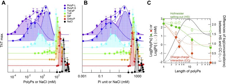
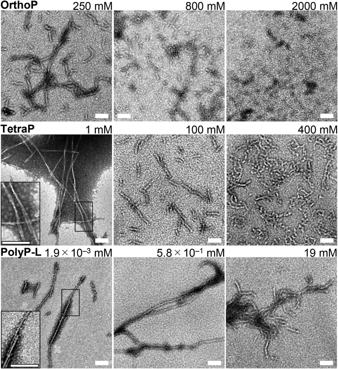
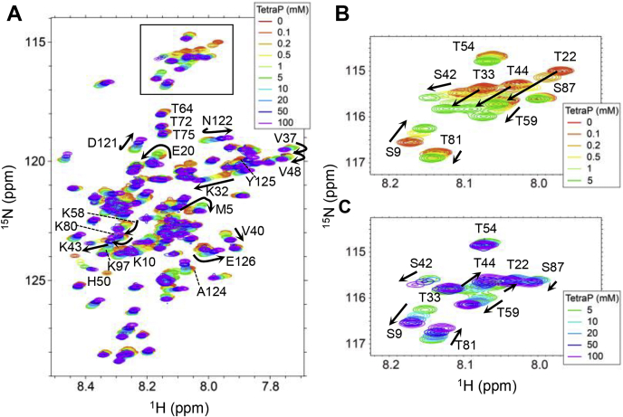
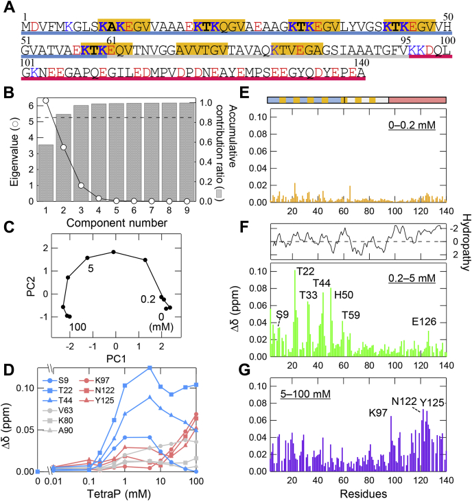
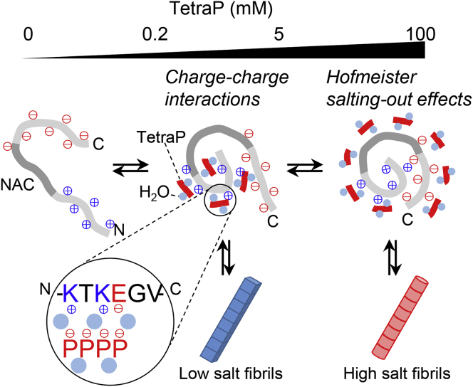
Polyphosphates (polyPs), chains of phosphate residues found in species across nature from bacteria to mammals, were recently reported to accelerate the amyloid fibril formation of many proteins. How polyPs facilitate this process, however, remains unknown. To gain insight into their mechanisms, we used various physicochemical approaches to examine the effects of polyPs of varying chain lengths on ultrasonication-dependent α-synuclein (α-syn) amyloid formation. Although orthophosphate and diphosphate exhibited a single optimal concentration of amyloid formation, triphosphate and longer-chain phosphates exhibited two optima, with the second at a concentration lower than that of orthophosphate or diphosphate. The second optimum decreased markedly as the polyP length increased. This suggested that although the optima at lower polyP concentrations were caused by interactions between negatively charged phosphate groups and the positive charges of α-syn, the optima at higher polyP concentrations were caused by the Hofmeister salting-out effects of phosphate groups, where the effects do not depend on the net charge. NMR titration experiments of α-syn with tetraphosphate combined with principal component analysis revealed that, at low tetraphosphate concentrations, negatively charged tetraphosphates interacted with positively charged "KTK" segments in four KTKEGV repeats located at the N-terminal region. At high concentrations, hydrated tetraphosphates affected the surface-exposed hydrophilic groups of compact α-syn. Taken together, our results suggest that long-chain polyPs consisting of 60 to 70 phosphates induce amyloid formation at sub-μM concentrations, which are comparable with the concentrations of polyPs in the blood or tissues. Thus, these findings may identify a role for polyPs in the pathogenesis of amyloid-related diseases.

Keywords: amyloid; biophysics; nuclear magnetic resonance (NMR); polyphosphate; protein aggregation; solubility; supersaturation; synuclein.


Conflict of interest statement

Conflicts of interest The authors declare that they have no conflicts of interest with the contents of this article.

Figures

Figure 1
Figure 1
Amyloid formation of α-syn in the presence of different kinds of polyPs.A, chemical structure of polyP. n is the degree of polymerization of the phosphate group. BD, kinetics of amyloid formation monitored by ThT fluorescence at varying concentrations of orthoP (B), tetraP (C), and polyP-L (D). The concentrations of polyPs are shown in the figures. EG, maximum values of ThT fluorescence (blue) and lag times (black) in the presence of orthoP (E), tetraP (F), and polyP-L (G). Each point and bar represent the average and standard deviation of five independent experiments, respectively. Maximum values of ThT fluorescence in Figures 1 and 2 were fitted with three Gaussian curves including the charge–charge interactions (orange), Hofmeister salting-out effects (green) and higher unknown component (gray).
Figure 2
Figure 2
PolyP concentration dependency of the amyloid formation of α-syn.A and B, amyloid formation in the presence of varying chain lengths and concentrations of polyPs plotted against polyP or NaCl concentrations (A), or phosphate unit or NaCl concentrations (B). Types of polyPs are shown in the figures. Data for orthoP, tetraP, and polyP-L are reproduced from Figure 1. C, logarithmic plots of optimal amyloid concentrations, which are caused by the charge–charge interactions (orange) and Hofmeister salting-out effects (green), obtained using the polyP concentration (closed circles), phosphate unit concentration (open circles), and NaCl concentration (closed triangles).
Figure 3
Figure 3
TEM images of α-syn aggregates in the presence of varying concentrations of polyPs.Top, orthoP, (middle) tetraP, and (bottom) polyP-L. The scale bars are 100 nm. Insets show the enlarged images of square regions.
Figure 4
Figure 4
1H-15N HSQC spectra of α-syn in the presence of varying concentrations of tetraP (A). Representative amino acid residues with a large shift are indicated in the figure. B and C, enlarged images of the enclosed area in the figure (A) at the concentrations of 0 to 5 (B) and 5 to 100 mM tetraP (C).
Figure 5
Figure 5
Interactions between tetraP and α-syn analyzed by NMR.A, amino acid sequence of α-syn, where the N-terminal, NAC, and C-terminal regions are underlined by blue, gray, and red, respectively, and “KTKEGV” repeat motifs are marked with orange. At pH 7.5, the positively and negatively charged residues are shown by blue and red, respectively. B and C, PCA of the NMR data against tetraP concentrations. B, the eigenvalues (circles) and accumulative contribution ratio (bars) of PCA. The dashed line indicates an accumulative contribution ratio of 0.85. C, the change in PC 1 and 2 scores depending on the tetraP concentrations. D, representative CSPs of HSQC peaks as a function of the molar concentration of tetraP. Colors correspond to the N-terminal (light blue), NAC (gray), and C-terminal (pink) regions, and their amino acid residues are shown in the figure. EG, the CSPs between 0 and 0.2 mM (E), 0.2 and 5 mM (F), and 5 and 100 mM (G). The bar above panel (E) shows the sequence motifs of α-syn, with the same colors as those used in (A). Panel (F) also shows the hydropathy plot of α-syn against the residue number (60).
Figure 6
Figure 6
Schematic of the mechanisms for polyP-induced amyloid formation of α-syn at pH 7.0. α-Syn with a pI of 4.7 has a net charge of −10 at pH 7.0. In the absence of tetraP or salts, the negative net charge causes the extended α-syn conformation, although the positive and negative charges are clustered on the N- and C-terminal regions, respectively. At the lower concentrations of tetraP, negatively charged tetraPs interact with positively charged “KTK” segments in KTKEGV repeat motifs via charge–charge interactions. Shielding of the charge repulsions leads to compact NAC regions stabilized by hydrophobic interactions and the formation of low-salt amyloid fibrils. At higher concentrations of tetraP, hydrated tetraPs cause Hofmeister salting-out effects and lead to the formation of high salt amyloid fibrils, in which tetraPs affect surface-exposed protein-charged groups of compact monomers.

Similar articles

Cited by

References

    1. Riek R., Eisenberg D.S. The activities of amyloids from a structural perspective. Nature. 2016;539:227–235. - PubMed
    1. Sipe J.D., Benson M.D., Buxbaum J.N., Ikeda S.I., Merlini G., Saraiva M.J., Westermark P. Amyloid fibril proteins and amyloidosis: Chemical identification and clinical classification International Society of Amyloidosis 2016 nomenclature guidelines. Amyloid. 2016;23:209–213. - PubMed
    1. Chiti F., Dobson C.M. Protein misfolding, amyloid formation, and human disease: A summary of progress over the last decade. Annu. Rev. Biochem. 2017;86:27–68. - PubMed
    1. Guerrero-Ferreira R., Taylor N.M., Mona D., Ringler P., Lauer M.E., Riek R., Britschgi M., Stahlberg H. Cryo-EM structure of alpha-synuclein fibrils. Elife. 2018;7 - PMC - PubMed
    1. Schweighauser M., Shi Y., Tarutani A., Kametani F., Murzin A.G., Ghetti B., Matsubara T., Tomita T., Ando T., Hasegawa K., Murayama S., Yoshida M., Hasegawa M., Scheres S.H.W., Goedert M. Structures of alpha-synuclein filaments from multiple system atrophy. Nature. 2020;585:464–469. - PMC - PubMed

Publication types

MeSH terms

LinkOut - more resources