. 2021 Jan-Jun:296:100512.
doi: 10.1016/j.jbc.2021.100512. Epub 2021 Mar 4.

The phosphorylation of the Smad2/3 linker region by nemo-like kinase regulates TGF-β signaling

Affiliations

The phosphorylation of the Smad2/3 linker region by nemo-like kinase regulates TGF-β signaling

Junbo Liang et al. J Biol Chem. 2021 Jan-Jun.

Abstract

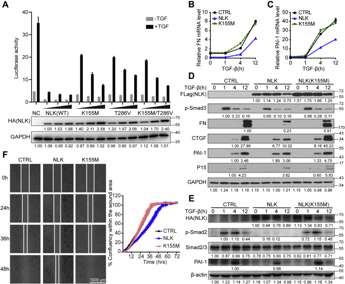
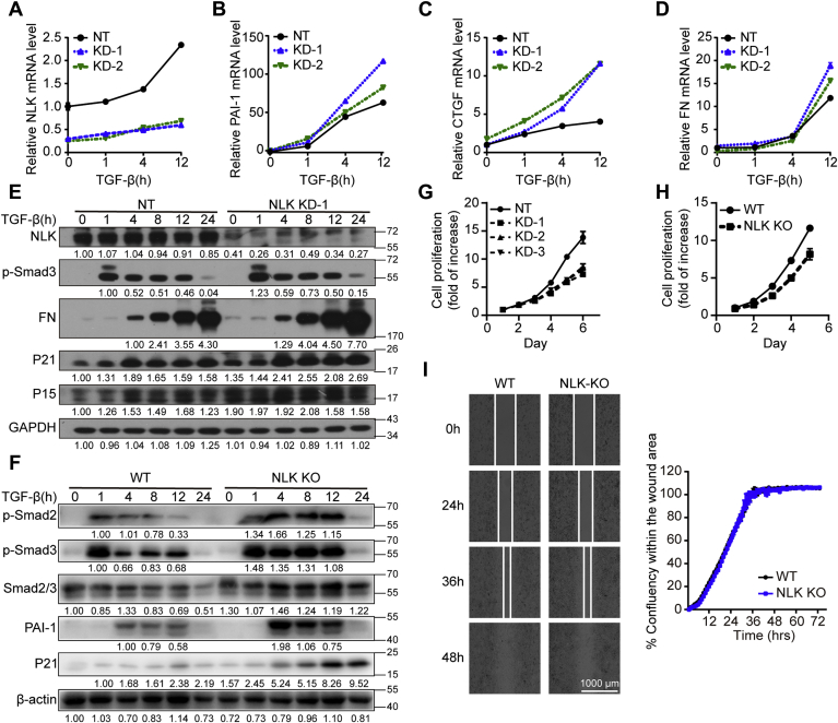
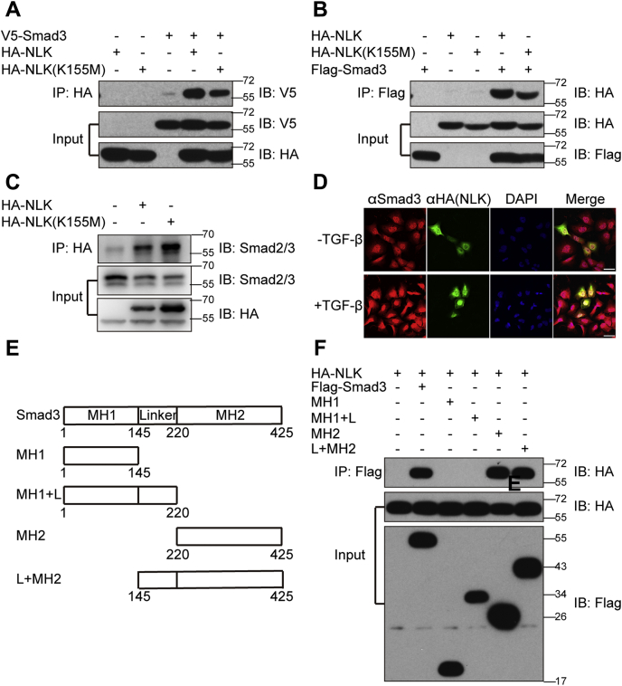
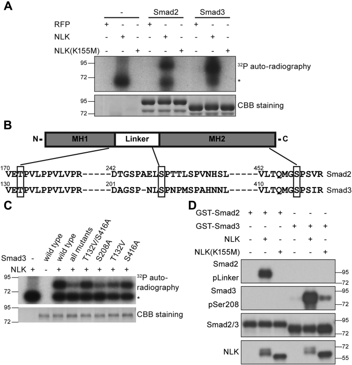
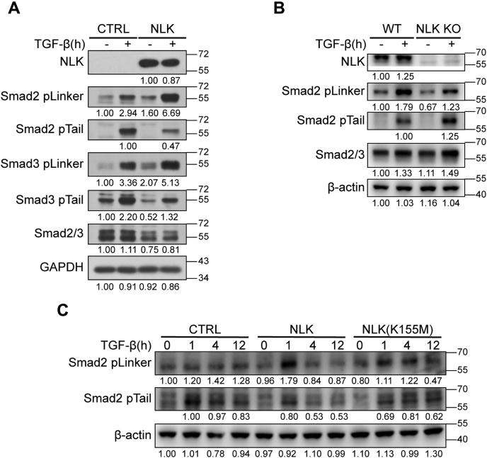
Smad2 and Smad3 (Smad2/3) are structurally similar proteins that primarily mediate the transforming growth factor-β (TGF-β) signaling responsible for driving cell proliferation, differentiation, and migration. The dynamics of the Smad2/3 phosphorylation provide the key mechanism for regulating the TGF-β signaling pathway, but the details surrounding this phosphorylation remain unclear. Here, using in vitro kinase assay coupled with mass spectrometry, we identified for the first time that nemo-like kinase (NLK) regulates TGF-β signaling via modulation of Smad2/3 phosphorylation in the linker region. TGF-β-mediated transcriptional and cellular responses are suppressed by NLK overexpression, whereas NLK depletion exerts opposite effects. Specifically, we discovered that NLK associates with Smad3 and phosphorylates the designated serine residues located in the linker region of Smad2 and Smad3, which inhibits phosphorylation at the C terminus, thereby decreasing the duration of TGF-β signaling. Overall, this work demonstrates that phosphorylation on the linker region of Smad2/3 by NLK counteracts the canonical phosphorylation in response to TGF-β signals, thus providing new insight into the mechanisms governing TGF-β signaling transduction.

Keywords: NLK; Smad2; Smad3; TGF-β signaling; linker phosphorylation.


Conflict of interest statement

Conflict of interest The authors declare that they have no conflict of interest.

Figures

Figure 1
Figure 1
NLK inhibits TGF-β-induced cell migration and transcriptional responses.A, NLK affects the SBE4-luc response. The SBE4-luc reporter was cotransfected with different doses (25 ng, 50 ng, and 100 ng) of NLK (WT) or its inactive kinase forms NLK(K155M), NLK(T286V), and NLK(K155M/T286V) in HEK293 cells treated with or without 100 pM TGF-β for 24 h. The data were measured in triplicate. Values and bars represent the means and the standard deviation. The protein levels of NLK were shown below, and the relative band intensity was shown under each panel (GAPDH: relative values of band intensity; HA: relative values of the band intensity ratio of target protein to GAPDH). B and C, quantitative real-time PCR (qRT-PCR) analysis of mRNA. NLK (stably expressing HA-NLK) cells, K155M (stably expressing HA-NLK(K155M)) cells, and parental HaCaT cells were stimulated with TGF-β at the indicated times before total RNA was extracted. The data were measured in triplicate. Values and bars represent the means and the standard deviation. D, TGF-β-induced expression of PAI-1, CTGF, FN, and P15 was inhibited by NLK. HaCaT cells stably expressing FLAG-NLK or FLAG-NLK K155M were stimulated with 100 pM TGF-β for the indicated times. The relative band intensity was shown under each panel (GAPDH: relative values of band intensity; others: relative values of the band intensity ratio of target protein to GAPDH). E, TGF-β-induced expression of C-tail phosphorylated Smad2, Smad2/3, and PAI-1 was inhibited by NLK. HeLa cells stably expressing HA-NLK or HA-NLK K155M were stimulated with 100 pM TGF-β for the indicated times. The relative band intensity was shown under each panel (β-actin: relative values of band intensity; others: relative values of the band intensity ratio of target protein to β-actin). F, HeLa cells prepared as in D were tested in a wound-healing assay. Wounds were performed by seeding cells into the Culture-Insert 2 Well. Cells were treated with 100 pM TGF-β and allowed to migrate for 72 h. The data were measured in triplicate. Values and bars represent the means and the standard deviation.
Figure 2
Figure 2
Depletion of NLK enhanced TGF-β-induced transcriptional responses and compromises cell growth.AD, qRT-PCR analysis of mRNA. HaCaT cells were transfected with NLK siRNA (KD-1 and KD-2) and nontargeting siRNA (NT), then stimulated with TGF-β at the indicated times before total RNA was extracted. The data were measured in triplicate. Values and bars represent the means and standard deviation values. E, western blot analysis of P21, P15, FN, and p-Smad3. Whole-cell lysates from HaCaT cells stably expressing NLK shRNA (NLK KD-1) and nontargeting siRNA (NT) were immunoblotted with antibodies as shown on the left. The relative band intensity was shown under each panel (GAPDH: relative values of band intensity; others: relative values of the band intensity ratio of target protein to GAPDH). F, western blot analysis of C-tail-phosphorylated Smad2/3, total Smad2/3, PAI-1, and P21 in the HeLa cells. Whole-cell lysates from the NLK-KO and WT cells were immunoblotted with antibodies as shown on the right. The relative band intensity was shown under each panel (β-actin: relative values of band intensity; others: relative values of the band intensity ratio of target protein to β-actin). G, proliferation of the HaCaT cells stably expressing NLK shRNA (KD-1; KD-2; KD-3) or nontargeting shRNA (NT) was examined by using the CCK-8 method. The data were measured in triplicate. Values and bars denote the means and standard deviation values. H, proliferation of NLK-knockout HeLa (NLK KO) cells or wild-type HeLa (WT) cells was examined by using the CCK-8 method. The data were measured in triplicate. Values and bars denote the means and standard deviation values. I, NLK-knockout HeLa (NLK KO) cells or wild-type HeLa (WT) cells were tested in a wound-healing assay. Wounds were performed by seeding cells into the Culture-Insert 2 Well. Cells were treated with 100 pM TGF-β and allowed to migrate for 72 h. The data were measured in triplicate. Values and bars represent the means and the standard deviation.
Figure 3
Figure 3
NLK interacted with Smad3.AC, co-immunoprecipitation of NLK with Smad2/3. HEK293T cells were transfected with expression plasmids as indicated. The immunoprecipitates were immunoblotted with the corresponding epitope antibodies. D, subcellular colocalization of NLK and Smad3 in the U2OS cells. U2OS cells were transfected with HA-tagged NLK for 24 h, treated with TGF-β for 1 h, and fixed. The localization of endogenous Smad3 was examined by indirect immunostaining with anti-Smad3 antibody (red). Cells treated with HA-NLK were observed through immunostaining with anti-HA antibody (green). E, schematic representation of Smad3 and its deletion mutants used in F. F, map of the region in Smad3 that interacts with NLK. Wild-type Smad3 and its deletion mutants were coexpressed with NLK in HEK293T cells. Immunoprecipitation and immunoblotting were performed as described in AC.
Figure 4
Figure 4
NLK phosphorylated Smad3 at Ser208 and Smad2 at the analogous site in vitro.A, NLK phosphorylated Smad2 and Smad3 in vitro. Flag-NLK, the catalytically inactive mutant Flag-NLK (K155M), or Flag-RFP immunoprecipitated was from HEK23T cells, and bacterially purified GST-Smad2 or GST-Smad3 was subjected to in vitro kinase reactions in the presence of [γ-32P] ATP. Samples were resolved by SDS-PAGE. Phosphorylated proteins were visualized by autoradiography. The asterisk denotes autophosphorylated NLK. B, schematic diagram of Smad2 and Smad3 phosphorylation sites as identified by mass spectrometry. GST-Smad2 and GST-Smad3 proteins phosphorylated by NLK were gathered form gel described in A and used in mass spectrometry analysis. C, the substitution of Ser208 with alanine in Smad3 abolished the phosphorylation of Smad3 by NLK. Bacterially purified GST-Smad3 and its point mutants were incubated with or without NLK in the presence of [γ-32P] ATP. Phosphorylated proteins were visualized by autoradiography. The asterisk denotes significant NLK autophosphorylation. CBB staining showed equalized Smad3 proteins in different reaction systems. D, an in vitro kinase assay was performed as described in A, and phosphorylated Smad2 and Smad3 were detected by the indicated phospho-specific antibodies. Equal substrate loading was confirmed by blotting with an anti-Smad2/3 antibody.
Figure 5
Figure 5
NLK is required for the Smad2/3 linker phosphorylation in cells.A, HaCaT stable cells as described in Figure 1D were stimulated with TGF-β for 1 h. Whole-cell lysates were prepared for western blot analysis with the indicated antibodies. The relative band intensity was shown under each panel (GAPDH: relative values of band intensity; others: relative values of the band intensity ratio of target protein to GAPDH). B, wild-type (WT) and NLK knockout (NLK KO) HeLa cells were stimulated with TGF-β for 1 h. Whole-cell lysates were prepared for western blot analysis with the indicated antibodies. The relative band intensity was shown under each panel (β-actin: relative values of band intensity; others: relative values of the band intensity ratio of target protein to β-actin). C, NLK knockout (NLK-KO) HeLa cells were infected with lentivirus expressing NLK or the NLK mutant (K155M), then stimulated with 100 pM TGF-β for the indicated times. Whole-cell lysates were prepared for western blot analysis with the indicated antibodies. The relative band intensity was shown under each panel (β-actin: relative values of band intensity; others: relative values of the band intensity ratio of target protein to β-actin).
Figure 6
Figure 6
The linker region phosphorylation destabilizes Smad2/3 and inhibits TGF-β signaling.A, validation of Smad2/3 KO in HeLa cells by western blot. B, validation of Smad2/3 KO in HeLa cells by Sanger sequencing. C and D, Smad2/3-KO HeLa cells were infected with lentivirus expressing wild-type or mutant Smad2/3, then treated with 100 μg/ml CHX for the indicated times. Whole-cell lysates were prepared for western blot analysis with the indicated antibodies. The relative band intensity was shown under each panel (β-actin: relative values of band intensity; Flag: relative values of the band intensity ratio of target protein to β-actin). E, NLK inhibits TGF-β signaling via the phosphorylation of the linker region. Smad2/3-deficient HeLa cells were infected with lentivirus expressing NLK and either Smad3(WT) or Smad3(S208A). HeLa cells were stimulated with 100 pM TGF-β for the indicated times. Subsequently, the total cell lysates were prepared for western blotting. The relative band intensity was shown under each panel (β-actin: relative values of band intensity; others: relative values of the band intensity ratio of target protein to β-actin).

Similar articles

Cited by

References

    1. Moses H.L., Roberts A.B., Derynck R. The discovery and early days of TGF-beta: A historical perspective. Cold Spring Harb. Perspect. Biol. 2016;8 - PMC - PubMed
    1. Yu Y., Feng X.H. TGF-beta signaling in cell fate control and cancer. Curr. Opin. Cell Biol. 2019;61:56–63. - PubMed
    1. Morikawa M., Derynck R., Miyazono K. TGF-beta and the TGF-beta family: Context-dependent roles in cell and tissue physiology. Cold Spring Harb. Perspect. Biol. 2016;8 - PMC - PubMed
    1. Derynck R., Budi E.H. Specificity, versatility, and control of TGF-beta family signaling. Sci. Signal. 2019;12 - PMC - PubMed
    1. Massague J. How cells read TGF-beta signals. Nat. Rev. Mol. Cell Biol. 2000;1:169–178. - PubMed

Publication types