. 2021 Mar:65:103255.
doi: 10.1016/j.ebiom.2021.103255. Epub 2021 Mar 4.

Camostat mesylate inhibits SARS-CoV-2 activation by TMPRSS2-related proteases and its metabolite GBPA exerts antiviral activity

Affiliations

Camostat mesylate inhibits SARS-CoV-2 activation by TMPRSS2-related proteases and its metabolite GBPA exerts antiviral activity

Markus Hoffmann et al. EBioMedicine. 2021 Mar.

Abstract

Background: Antivirals are needed to combat the COVID-19 pandemic, which is caused by SARS-CoV-2. The clinically-proven protease inhibitor Camostat mesylate inhibits SARS-CoV-2 infection by blocking the virus-activating host cell protease TMPRSS2. However, antiviral activity of Camostat mesylate metabolites and potential viral resistance have not been analyzed. Moreover, antiviral activity of Camostat mesylate in human lung tissue remains to be demonstrated.

Methods: We used recombinant TMPRSS2, reporter particles bearing the spike protein of SARS-CoV-2 or authentic SARS-CoV-2 to assess inhibition of TMPRSS2 and viral entry, respectively, by Camostat mesylate and its metabolite GBPA.

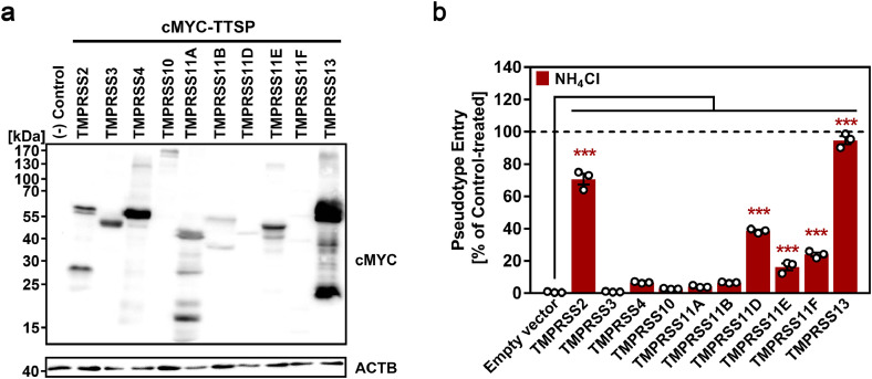
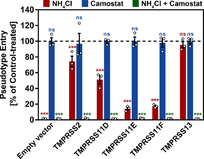
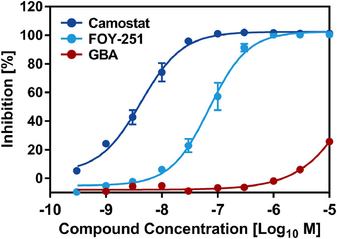
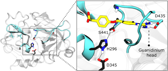
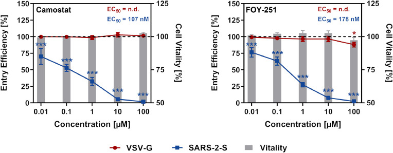
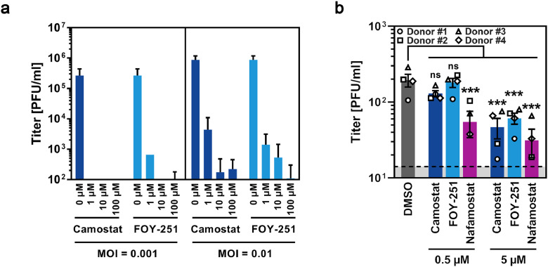
Findings: We show that several TMPRSS2-related proteases activate SARS-CoV-2 and that two, TMPRSS11D and TMPRSS13, are robustly expressed in the upper respiratory tract. However, entry mediated by these proteases was blocked by Camostat mesylate. The Camostat metabolite GBPA inhibited recombinant TMPRSS2 with reduced efficiency as compared to Camostat mesylate. In contrast, both inhibitors exhibited similar antiviral activity and this correlated with the rapid conversion of Camostat mesylate into GBPA in the presence of serum. Finally, Camostat mesylate and GBPA blocked SARS-CoV-2 spread in human lung tissue ex vivo and the related protease inhibitor Nafamostat mesylate exerted augmented antiviral activity.

Interpretation: Our results suggest that SARS-CoV-2 can use TMPRSS2 and closely related proteases for spread in the upper respiratory tract and that spread in the human lung can be blocked by Camostat mesylate and its metabolite GBPA.

Funding: NIH, Damon Runyon Foundation, ACS, NYCT, DFG, EU, Berlin Mathematics center MATH+, BMBF, Lower Saxony, Lundbeck Foundation, Novo Nordisk Foundation.

Keywords: Camostat; FOY-251; GBPA; SARS-CoV-2; TMPRSS2.


Conflict of interest statement

Declaration of Competing Interests H.H-W., N.K., L.K.S., O.S.S., J.B.H., M.W., S.O., O.D., D.J., S.L., F.N., M.K. have nothing to disclose. T.Y., K.Y., H.M. report personal fees from Ono Pharmaceutical, during the conduct of the study. M.H. reports grants from Deutsche Forschungsgemeinschaft (DFG, German Research Foundation), during the conduct of the study. J.C.S. reports personal fees from Google, personal fees from Meliora Therapeutics, outside the submitted work. J.C.S. is an employee of Google. This work was performed outside of her affiliation with Google and used no proprietary knowledge or materials from Google. P.A. reports grants from Country of Lower Saxony, during the conduct of the study. T. H. reports grants from Deutsche Forschungsgemeinschaft (DFG) SFB/TRR 186, during the conduct of the study. L.R. reports grants from Bayer AG, outside the submitted work. A.B. reports grants from Fraunhofer DRECOR (Drug Repurposing for Corona), during the conduct of the study; other from Fraunhofer ITEM, outside the submitted work. J.M.S. reports grants from NIH, grants from New York Community Trust, grants from Damon Runyon Foundation, grants from American Cancer Society, grants from Department of Defense, during the conduct of the study; personal fees from Meliora Therapeutics, personal fees from Tyra Biosciences, personal fees from Ono Pharmaceutical, outside the submitted work. S.P. reports grants from Bundesministerium für Bildung und Forschung, grants from Deutsche Forschungsgemeinschaft, grants from Country of Lower Saxony, during the conduct of the study; other from Ono Pharmaceutical, outside the submitted work.

Figures

Fig 1
Fig. 1
Different type-II transmembrane serine proteases (TTSPs) can activate SARS-2-S in transfected cells. (a) The indicated TTSPs equipped with an N-terminal C-MYC antigenic tag were transiently expressed in 293T cells and expression analyzed by immunoblot with anti-C-MYC antibody. Detection of ß-actin (ACTB) served as loading control. Similar results were obtained in three biological replicates. (b) BHK-21 cells transiently expressing ACE2 and one of the indicated TTSPs (or empty vector) were pre-incubated with either 50 mM ammonium chloride or DMSO (control, indicated by dashed line) for 2 h, before they were inoculated with particles pseudotyped with SARS-2-S. At 16 h post inoculation, SARS-2-S-driven cell entry of viral pseudotypes was analyzed by measuring the activity of virus-encoded luciferase activity in cell lysates. Data were further normalized and entry efficiency in the absence of ammonium chloride was set as 100 % (indicated by dashed line). Shown are the average (mean) data obtained from three biological replicates, each performed with four technical replicates. Error bars indicate the standard error of the mean (SEM). Statistical significance of differences in entry efficiency in the presence of ammonium chloride was analyzed by two-way analysis of variance (ANOVA) with Dunnett's posttest (P values, from left to right: 0.0001; 0.9999; 0.0620; 0.9140; 0.4900; 0.0685; 0.0001; 0.0001; 0.0001; 0.0001).
Fig 2
Fig. 2
SARS-2-S activating proteases are expressed in lung and blood. (a) T-SNE clustering of cells from the human lung . Cells expressing the coronavirus receptor ACE2 are highlighted in the right panel. These panels are reproduced with permission from Smith et al. . Cells expressing various S-activating proteases in the human lung are highlighted. (b) T-SNE clustering of cells from the human airway . Cells expressing the coronavirus receptor ACE2 and the various S-activating proteases in the human airway are highlighted. (c) Log2-normalized expression data of the indicated genes across different human tissues from the GTEx consortium . Certain panels are reprinted from “Cigarette Smoke Exposure and Inflammatory Signaling Increase the Expression of the SARS-CoV-2 Receptor ACE2 in the Respiratory Tract”, Developmental Cell 53, Joan C. Smith, Erin L. Sausville, Vishruth Girish, Monet Lou Yuan, Anand Vasudevan, Kristen M. John, Jason M. Sheltzer, 514–529, Copyright (2020), with permission from Elsevier.
Fig 3
Fig. 3
Activation of SARS-2-S by TMPRSS2-related proteases can be suppressed by Camostat mesylate. The experiment was performed as described for Fig. 1 with the modifications that only TMPRSS2, TMPRSS11D, TMPRSS11E, TMPRSS11F and TMPRSS13 were investigated and target cells were pre-treated with either 50 mM ammonium chloride (red), 100 µM Camostat mesylate (blue) or a combination of both (green). DMSO-treated cells served as controls. At 16 h post inoculation with viral particles bearing SARS-2-S, pseudotype entry was analyzed by measuring virus-encoded luciferase activity in cell lysates. Data were further normalized and entry efficiency into control-treated cells was set as 100%. Shown are the average (mean) data obtained from three biological replicates, each performed with four technical replicates. Error bars indicate the SEM. Statistical significance of differences in entry efficiency in ammonium chloride-, Camostat mesylate- or ammonium chloride + Camostat mesylate-treated cells versus control-treated cells was analyzed by two-way ANOVA with Dunnett's posttest (P values, from left to right: NH4Cl [0.0001; 0.0001; 0.0001; 0.0001; 0.0001; 0.7334]; Camostat [0.9999; 0.8995; 0.9969; 0.9999; 0.9731; 0.9999]; NH4Cl/Camostat [0.0001; 0.0001; 0.0001; 0.0001; 0.0001; 0.0001]).
Fig 4
Fig. 4
Camostat mesylate and FOY-251 inhibit the activity of recombinant TMPRSS2. Incubation of recombinant TMPRSS2 with the Boc-Gln-Ala-Arg-MCA peptide substrate leads to the cleavage of the substrate and the release of the AMC(7-Amino-4-methylcoumarin) fluorophore, resulting in a fluorescent signal. Data were normalized against the fluorescence signals obtained in the absence of test compounds (Camostat mesylate, FOY-251, GBA). The concentration-response data for each test compound were plotted and modeled by a four-parameter logistic fit to determine the 50% effective concentration (EC50) value. Inhibitory activity of Camostat mesylate (blue), FOY-251(light blue) and GBA (red) against TMPRSS2 recombinant protein were visualized and curve fitting was performed using GraphPad Prism. The average of two biological replicates, each performed with four (Camostat mesylate and FOY-251) or two technical replicates (GBA) is shown. EC50 values were 4 nM (Camostat mesylate), 70 nM (FOY-251), >10 µM (GBA).
Fig 5
Fig. 5
TMPRSS2 protease domain and GBPA interaction. A TMPRSS2 structure model is shown in the left panel, the active site is highlighted in cyan and catalytic triad residues are shown in black. The representative structure of GBPA bound to TMPRSS2 in a reactive complex is shown in the right panel. The GBPA guanidinium head forms a salt bridge with Asp-435 inside the S1 pocket. This transient complex, which is similar for Camostat, is prone to be catalyzed at the ester bond interacting with Ser-441, leading to a covalent complex with TMPRSS2 inhibited.
Fig 6
Fig. 6
Camostat mesylate is rapidly converted into GBPA in the presence of cell culture medium. (a) Metabolization of Camostat mesylate. (b) LC-MS/MS determination of Camostat, GBPA and GBA in culture medium containing FCS. Camostat mesylate was added to FCS-containing culture medium at a concentration of 15 µM. Samples were taken after incubation for 1, 15, 30, 60, 120, 240, 480, and 1,440 min at 37 °C, snap-frozen and stored at -80 °C. Samples were analyzed by LC-MS/MS and quantified regarding their content of intact Camostat mesylate and its metabolites GBPA (active) and GBA (inactive). Presented are the average (mean) data from three biological replicates (each performed with single samples). Error bars indicate the SEM. The turnover time that is required to cause metabolization of 50 % of Camostat mesylate (T1/2) was further calculated by a non-linear regression model and was determined to be 141.3 min (95% confidence interval = 116.5 to 171.7 min). (c) Relative levels of Camostat mesylate and GBPA after incubation of 15 µM Camostat mesylate in either water or FCS-containing culture medium. For normalization, the combined values of Camostat mesylate and GBPA were set as 100% and the relative fractions of the compounds were calculated. Presented are the average (mean) data from three biological replicates (each performed with single samples). Error bars indicate SEM. Statistical significance of differences in GBPA levels following incubation of Camostat mesylate in either water or FCS-containing culture medium was analyzed by paired, two-tailed student's t-test (P = 0.0090). Abbreviations: FOY-51/GBPA = 4-(4-guanidinobenzoyloxy)phenylacetic acid; GBA = 4-guanidinobenzoic acid.
Fig 7
Fig. 7
Camostat mesylate and FOY-251 inhibit SARS-2-S-driven cell entry with comparable efficiency. Calu-3 cells were pre-incubated with different concentrations of Camostat mesylate (left panel), FOY-251 (right panel) or DMSO (control, indicated by dashed lines) for 2 h, before being inoculated with pseudotype particles bearing VSV-G (red) or SARS-2-S (blue). Alternatively, in order to analyze potential negative effects of Camostat mesylate and FOY-251 on cell vitality (grey bars), cells received medium instead of pseudotype particles and were further incubated. At 16 h post inoculation, pseudotype entry and cell vitality were analyzed by measuring the activity of virus-encoded luciferase in cell lysates or intracellular adenosine triphosphate levels (CellTiter-Glo assay), respectively. Data were further normalized and entry efficiency/cell vitality in the absence of Camostat mesylate and FOY-251 was set as 100%. Shown are the average (mean) data obtained from three biological replicates, each performed with four technical replicates. Error bars indicate SEM. Statistical significance of differences in entry efficiency/cell vitality in Camostat mesylate - or FOY-251-treated cells versus control-treated cells was analyzed by two-way ANOVA with Dunnett's posttest (P values, from left to right: Camostat/VSV-G [0.9999; 0.9999; 0.9996; 0.9733; 0.9986]; Camostat/SARS-2-S [0.0001; 0.0001; 0.0001; 0.0001; 0.0001]; Camostat/Cell vitality [0.9999; 0.9999; 0.9999; 0.9998; 0.9810]; FOY-251/VSV-G [0.9997; 0.9730; 0.8867; 0.8838; 0.0326]; FOY-251/SARS-2-S [0.0001; 0.0001; 0.0001; 0.0001; 0.0001]; FOY-251/Cell vitality [0.9986; 0.9765; 0.9455; 0.9460; 0.9612]).
Fig 8
Fig. 8
Camostat mesylate and FOY-251 inhibit SARS-CoV-2 infection with comparable efficiency. (a) Calu-3 cells were pre-incubated for 2h with double concentration of Camostat mesylate or FOY-251 as indicated. DMSO-treated cells served as control. Thereafter, cells were infected with SARS-CoV-2. After 1 h of incubation, the inoculum was removed and cells were washed with PBS, before culture medium containing inhibitor or DMSO was added. Culture supernatants were harvested at 24 h post infection and subjected to standard plaque formation assay. Viral titers were determined as plaque forming units per ml (pfu/ml). Presented are the data from a single experiment performed with technical triplicates and the results were confirmed in a separate experiment with another SARS-CoV-2 isolate. Error bars indicate the standard deviation. (b) Precision-cut lung slices (PCLS) from four donors were pre-treated with 0.5 or 5 µM Camostat mesylate, FOY-251 or Nafamostat mesylate before being inoculated with SARS-CoV-2. DMSO-treated PCLS served as control. After 1 h of incubation, the inoculum was removed and PCLS were washed with PBS, before culture medium containing the respective inhibitor (or DMSO) was added. Culture supernatants were harvested at 24 h post infection and subjected to standard plaque formation assay. Presented are the data from four biological replicates, each performed with four (donor 1) or three (donor 2-4) technical replicates. Error bars indicate the SEM. One-way ANOVA with Dunnett's posttest was used for statistical analysis. P values, from left to right: 0.5 µM Camostat [0.1551]; 0.5 µM FOY-251 [0.9919]; 0.5 µM Nafamostat [0.0006]; 5 µM Camostat [0.0003]; 5 µM FOY-251 [0.0010]; 5 µM Nafamostat [0.0001]).

Update of

Comment in

References

    1. World Health Organization (WHO) Coronavirus disease (COVID-19) Situation Report. 2020;184
    1. Santos J, Brierley S, Gandhi MJ, Cohen MA, Moschella PC, Declan ABL. Repurposing therapeutics for potential treatment of SARS-CoV-2: a review. Viruses. 2020;12(7) - PMC - PubMed
    1. Beigel JH, Tomashek KM, Dodd LE. Remdesivir for the treatment of COVID-19 - preliminary report. N Engl J Med. 2020 - PubMed
    1. Wang Y, Zhang D, Du G. Remdesivir in adults with severe COVID-19: a randomised, double-blind, placebo-controlled, multicentre trial. Lancet. 2020;395(10236):1569–1578. - PMC - PubMed
    1. Hoffmann M, Kleine-Weber H, Schroeder S. SARS-CoV-2 cell entry depends on ACE2 and TMPRSS2 and is blocked by a clinically proven protease inhibitor. Cell. 2020;181(2):271–280. e8. - PMC - PubMed

MeSH terms