. 2021 Apr 6;49(6):3308-3321.
doi: 10.1093/nar/gkab112.

Spatial coupling between DNA replication and mismatch repair in Caulobacter crescentus

Affiliations

Spatial coupling between DNA replication and mismatch repair in Caulobacter crescentus

Tiancong Chai et al. Nucleic Acids Res. .

Abstract

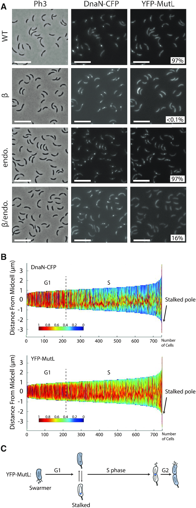
The DNA mismatch repair (MMR) process detects and corrects replication errors in organisms ranging from bacteria to humans. In most bacteria, it is initiated by MutS detecting mismatches and MutL nicking the mismatch-containing DNA strand. Here, we show that MMR reduces the appearance of rifampicin resistances more than a 100-fold in the Caulobacter crescentus Alphaproteobacterium. Using fluorescently-tagged and functional MutS and MutL proteins, live cell microscopy experiments showed that MutS is usually associated with the replisome during the whole S-phase of the C. crescentus cell cycle, while MutL molecules may display a more dynamic association with the replisome. Thus, MMR components appear to use a 1D-scanning mode to search for rare mismatches, although the spatial association between MutS and the replisome is dispensible under standard growth conditions. Conversely, the spatial association of MutL with the replisome appears as critical for MMR in C. crescentus, suggesting a model where the β-sliding clamp licences the endonuclease activity of MutL right behind the replication fork where mismatches are generated. The spatial association between MMR and replisome components may also play a role in speeding up MMR and/or in recognizing which strand needs to be repaired in a variety of Alphaproteobacteria.


Figures

Graphical Abstract
Graphical Abstract
In Caulobacter crescentus, MutS is recruited to the replication fork to facilitate detection of replication errors. The MutL endonuclease is licensed by the replisome when MutS detects a mismatch.
Figure 1.
Figure 1.
Comparison of the spontaneous mutation rates of different C. crescentus strains. This figure is based on values described in Supplementary Table S4. Relevant genotypes (and strain numbers) are indicated on the left side of the figure. To facilitate comparisons, values were normalized so that the value for a wild-type NA1000 strain equals 1. The spontaneous mutation rate of each strain was estimated by measuring the spontaneous appearance of rifampicin-resistant clones. Each value was estimated from minimum three independent cultures (standard deviations are described in Supplementary Table S4).
Figure 2.
Figure 2.
YFP-MutS forms discrete fluorescent foci in a majority of C. crescentus cells. The subcellular localization of several derivatives of YFP-MutS was analyzed in ΔmutS cells. Strains JC1433 (ΔmutS Pxyl::YFP-mutS) (A), JC1770 (ΔmutS Pxyl::YFP-mutS(849AAAAA853)) (B), JC1666 (ΔmutS Pxyl::YFP-mutS(F44A)) (C), JC1665 (ΔmutS Pxyl::YFP-mutS(K661M)) (D) and JC1739 (ΔmutS Pxyl::YFP-mutS(E735A)) (E) were cultivated into PYE medium and then transferred into M2G medium. 0.3% xylose was added to cultures when they reached an OD660 nm∼0.3. Cells were then imaged by fluorescence microscopy when the OD660 nm reached ∼0.5. Representative images are shown here. Ph3 indicates phase-contrast images. The % indicated onto images corresponds to the average proportion of cells (using values obtained from three independent experiments) displaying a distinct fluorescent focus (intensity >2-fold above background). The white scale bar corresponds to 8 μm.
Figure 3.
Figure 3.
YFP-MutS forms discrete fluorescent foci throughout the S-phase of the C. crescentus cell cycle. (A) Demograph showing the subcellular localization of YFP-MutS in ΔmutS cells sorted as a function of their size. JC1433 (ΔmutS Pxyl::YFP-mutS) cells were cultivated and imaged as described for Figure 2. Short cells correspond to G1/swarmer cells, while intermediate and longer cells correspond to stalked and pre-divisional S-phase cells, respectively. (B) Time-lapse fluorescence microscopy experiment showing the cell cycle localization of YFP-MutS as a function of the cell cycle of ΔmutS cells. JC1433 cells were first cultivated in PYE medium overnight and then diluted in M2G medium until the cells reached pre-exponential phase (OD660 nm = 0.1–0.2). Xylose at 0.3% was added into the cultures to induce the Pxyl promoter for 2.5 h. Swarmer cells were then isolated (synchronization protocol) from the cell culture, immobilized onto an agarose pad and imaged by fluorescence microscopy every 20 min. Representative images are shown here. The schematics drawn under the microscopy images highlight in red the subcellular localization of YFP-MutS in cells imaged above. (C) This schematic shows the C. crescentus cell cycle and the blue color highlights where MutS appears to be localized as a function of the cell cycle using results from panels A and B. This localization pattern is reminiscent of the known localization pattern of replisome components in C. crescentus (22).
Figure 4.
Figure 4.
YFP-MutS and YFP-MutL form frequent foci in C. crescentus cells, independently of mismatch frequency. (A) YFP-MutS localization in cells with a wild-type (WT) or a proofreading-deficient (dnaQ(G13E)) replicative DNA polymerase. Strains JC1433 (ΔmutS Pxyl::YFP-mutS) and JC1724 (ΔmutS Pxyl::YFP-mutS dnaQ(G13E)) were cultivated into PYE medium and then transferred into M2G medium. 0.3% xylose was added to cultures when they reached an OD660 nm∼0.3. Cells were then imaged by fluorescence microscopy when the OD660nm reached ∼0.5. (B) YFP-MutL localization in cells with a wild-type or a proofreading-deficient replicative DNA polymerase. Strains JC1825 (ΔmutL Pxyl::YFP-mutL), and JC1845 (ΔmutL Pxyl::YFP-mutL dnaQ(G13E)) were cultivated and imaged as described for panel A. Representative images are shown in panels A and B. Ph3 indicates phase-contrast images. The % indicated onto images corresponds to the average proportion of cells (using values obtained from three independent experiments) displaying a distinct fluorescent focus (intensity >2-fold above background). The white scale bar corresponds to 8μm.
Figure 5.
Figure 5.
YFP-MutL forms discrete fluorescent foci in a subset of S-phase C. crescentus cells. (A) Subcellular localization of several derivatives of YFP-MutL in ΔmutL cells. Strains JC1825 (ΔmutL Pxyl::YFP-mutL) labeled ‘WT’, JC1749 (ΔmutL Pxyl::YFP-mutL(497ATLAAP502)) labeled ‘β' and JC1667 (ΔmutL Pxyl::YFP-mutL(D472N)) labeled ‘endo’ were cultivated into PYE medium and then transferred into M2G medium. 0.3% xylose was added to cultures when they reached an OD660 nm∼0.3. Cells were then imaged by fluorescence microscopy when the OD660nm reached ∼0.5. The % indicated onto images corresponds to the average proportion of cells (using values obtained from three independent experiments) displaying a distinct fluorescent focus (intensity >2-fold above background). The white scale bar corresponds to 8 μm. (B) Time-lapse fluorescence microscopy experiment showing the localization of YFP-MutL as a function of the cell cycle of ΔmutL cells. JC1825 cells were first cultivated in PYE medium overnight and then diluted in M2G medium until the cells reached pre-exponential phase (OD660 nm = 0.1–0.2). Xylose at 0.3% was added into the cultures to induce the Pxyl promoter for 2.5 h. Swarmer cells were then isolated (synchronization protocol) from the cell culture, immobilized onto an agarose pad and imaged by fluorescence microscopy every 20 min. The schematics drawn under the microscopy images highlight the localization of YFP-MutL in cells imaged above. Representative images of cells are shown and Ph3 indicates phase-contrast images in panels A and B.
Figure 6.
Figure 6.
YFP-MutL foci co-localize with the replisome. (A) Subcellular localization of DnaN-CFP and of several derivatives of YFP-MutL in ΔmutL cells. Strains JC1812 (dnaN-CFP ΔmutL Pxyl::YFP-mutL) labeled ‘WT’, JC1750 (dnaN-CFP ΔmutL Pxyl::YFP-mutL(497ATLAAP502)) labeled ‘β", JC1670 (dnaN-CFP ΔmutL Pxyl::YFP-mutL(D472N)) labeled ‘endo’ and JC1753 (dnaN-CFP ΔmutL Pxyl::YFP-mutL(D472N -497ATLAAP502)) labeled ‘β/endo’, were cultivated into PYE medium and then transferred into M2G medium. 0.3% xylose was added to cultures when they reached an OD660nm∼0.3. Cells were then imaged by fluorescence microscopy when the OD660 nm reached ∼0.5. The % indicated onto images corresponds to the average proportion of distinct MutL-YFP foci (intensity >2-fold above average background) that are co-localized with DnaN-CFP foci (using values obtained from three independent experiments). The white scale bar corresponds to 8 μm. (B) Demographs showing the subcellular localization of DnaN-CFP and YFP-MutL in ΔmutL cells sorted as a function of their size. Strain JC1812 was cultivated and imaged as described for panel A. Short cells correspond to G1/swarmer cells, while intermediate and longer cells correspond to stalked and pre-divisional S-phase cells, respectively. (C) This schematic shows the C. crescentus cell cycle and the blue color highlights where YFP-MutL is localized as a function of the cell cycle based on images shown in panel B and in Figure 5B.
Figure 7.
Figure 7.
YFP-UvrD forms rare fluorescent foci in C. crescentus cells. Subcellular localization of YFP-UvrD in wild-type, dnaQ(G13E) or ΔmutS cells. Strains JC1946 (Pxyl::YFP-uvrD), JC2211 (dnaQ(G13E) Pxyl::YFP-uvrD) and JC1977 (ΔmutS Pxyl::YFP-uvrD) were cultivated into PYE medium and then transferred into M2G medium. 0.3% xylose was added to cultures when they reached an OD660 nm∼0.3. Cells were then imaged by fluorescence microscopy when the OD660nm reached ∼0.5. Representative images are shown here. Ph3 indicates phase-contrast images. The % indicated onto images corresponds to the average proportion of cells (using values obtained from three independent experiments) displaying a fluorescent focus (intensity >2-fold above background). The white scale bar corresponds to 11μm.
Figure 8.
Figure 8.
Model for the MMR process in C. crescentus. MutS-ADP binds to the β-clamp for 1D mismatch scanning during DNA replication. Mismatch detection by MutS triggers an ADP to ATP exchange and a conformational change in MutS, converting it into a sliding clamp that activates downstream MMR events. The ATP bound to MutS is then hydrolyzed, regenerating MutS-ADP that rapidly goes back to the replisome. MutL is dynamically recruited to the β-clamp during DNA replication and this interaction is needed for its activity as an endonuclease that nicks newly synthesized DNA strands. Mismatch detection by MutS most likely activates the latent endonuclease activity of MutL and/or helicases/exonucleases (Exo) needed for downstream events of the MMR process. The DNA polymerase III then resynthesizes the gap, while the ligase restores strand continuity.

Similar articles

Cited by

References

    1. Iyer R.R., Pluciennik A., Burdett V., Modrich P.L.. DNA mismatch repair: functions and mechanisms. Chem. Rev. 2006; 106:302–323. - PubMed
    1. Fishel R., Lescoe M.K., Rao M.R., Copeland N.G., Jenkins N.A., Garber J., Kane M., Kolodner R.. The human mutator gene homolog MSH2 and its association with hereditary nonpolyposis colon cancer. Cell. 1993; 75:1027–1038. - PubMed
    1. Gutierrez A., Laureti L., Crussard S., Abida H., Rodriguez-Rojas A., Blazquez J., Baharoglu Z., Mazel D., Darfeuille F., Vogel J.et al. .. beta-Lactam antibiotics promote bacterial mutagenesis via an RpoS-mediated reduction in replication fidelity. Nat. Commun. 2013; 4:1610. - PMC - PubMed
    1. Li G.M. Mechanisms and functions of DNA mismatch repair. Cell Res. 2008; 18:85–98. - PubMed
    1. Jiricny J. Postreplicative mismatch repair. Cold Spring Harb. Perspect. Biol. 2013; 5:a012633. - PMC - PubMed

Publication types

MeSH terms

LinkOut - more resources