. 2021 Mar 31;217(3):iyab006.
doi: 10.1093/genetics/iyab006.

An expanded auxin-inducible degron toolkit for Caenorhabditis elegans

Affiliations

An expanded auxin-inducible degron toolkit for Caenorhabditis elegans

Guinevere E Ashley et al. Genetics. .

Abstract

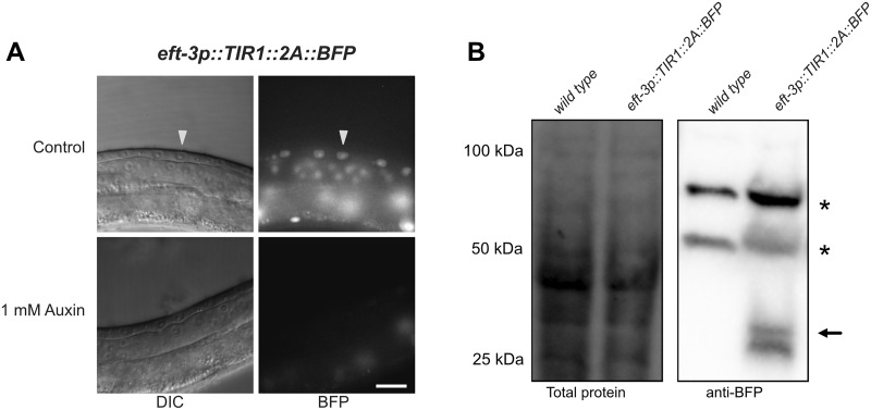
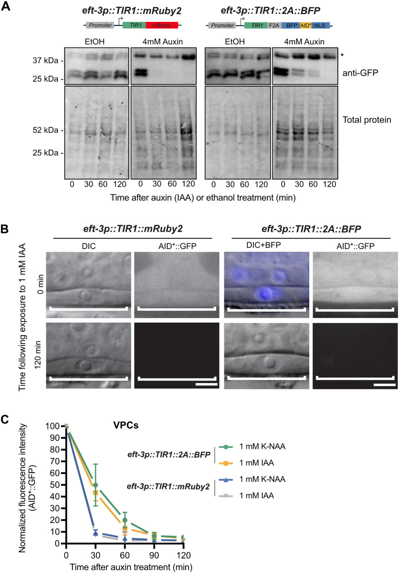
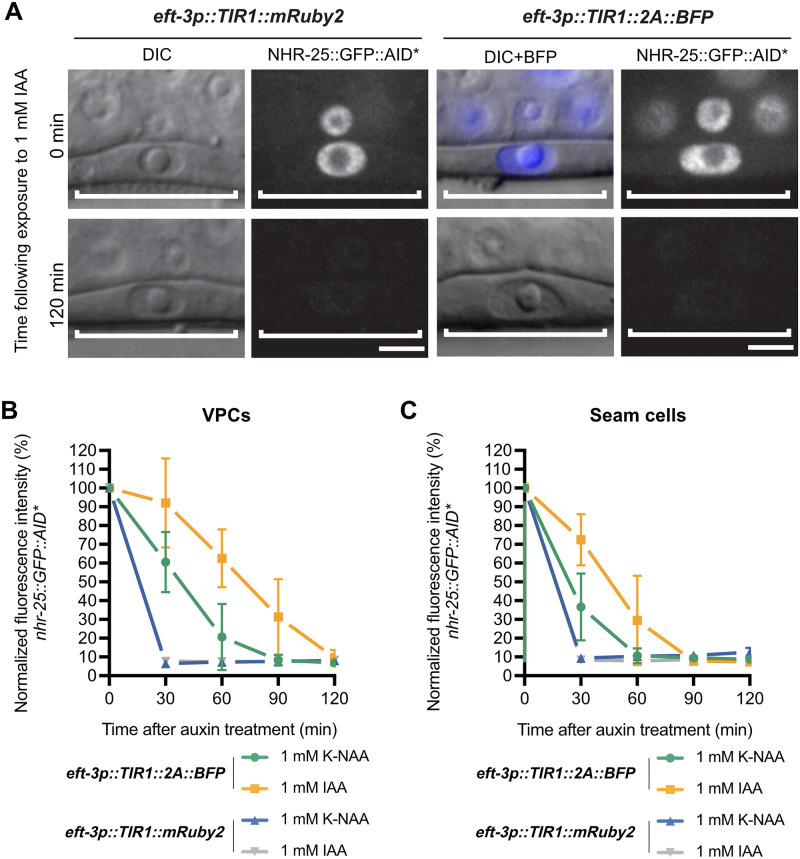
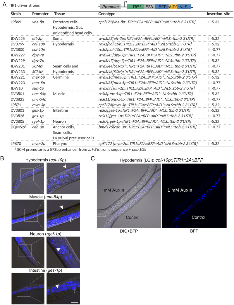
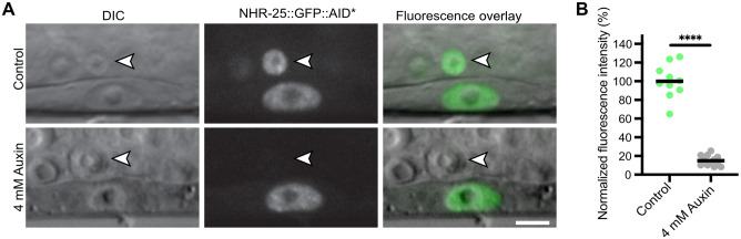
The auxin-inducible degron (AID) system has emerged as a powerful tool to conditionally deplete proteins in a range of organisms and cell types. Here, we describe a toolkit to augment the use of the AID system in Caenorhabditis elegans. We have generated a set of single-copy, tissue-specific (germline, intestine, neuron, muscle, pharynx, hypodermis, seam cell, anchor cell) and pan-somatic TIR1-expressing strains carrying a co-expressed blue fluorescent reporter to enable use of both red and green channels in experiments. These transgenes are inserted into commonly used, well-characterized genetic loci. We confirmed that our TIR1-expressing strains produce the expected depletion phenotype for several nuclear and cytoplasmic AID-tagged endogenous substrates. We have also constructed a set of plasmids for constructing repair templates to generate fluorescent protein::AID fusions through CRISPR/Cas9-mediated genome editing. These plasmids are compatible with commonly used genome editing approaches in the C. elegans community (Gibson or SapTrap assembly of plasmid repair templates or PCR-derived linear repair templates). Together these reagents will complement existing TIR1 strains and facilitate rapid and high-throughput fluorescent protein::AID tagging of genes. This battery of new TIR1-expressing strains and modular, efficient cloning vectors serves as a platform for straightforward assembly of CRISPR/Cas9 repair templates for conditional protein depletion.

Keywords: C. elegans; AID system; CRISPR/Cas9; SapTrap; Transport Inhibitor Response 1; self-excising cassette.


Figures

Figure 1
Figure 1
Schematic of the auxin-inducible degron (AID) system. The plant F-box protein TIR1 is expressed using a promoter of interest with a desired spatiotemporal expression pattern. TIR1 interacts with endogenous Skp1 and Cul1 proteins to form an SCF E3 ubiquitin ligase complex. An auxin-inducible degron sequence (AID) is fused to a protein of interest. In the presence of the plant hormone auxin, TIR1 recognizes and binds the AID sequence, leading to ubiquitination and subsequent degradation of the AID-tagged protein. We use a minimal, 44 amino acid degron sequence (AID*), but a full-length 229 amino acid AID tag or a 68 amino acid mini-AID (mAID) are used in other systems. In C. elegans, the system is frequently used with single-copy TIR1 transgenes inserted into neutral loci, and AID* knock-ins into genes of interest, though extrachromosomal arrays can also be used. Created with BioRender.com.
Figure 2
Figure 2
A new TIR1 expression system allows assessment of TIR1 expression and activity. (A) The new TIR1 expression construct contains a TIR1::F2A::BFP::AID*::NLS transgene cassette. An F2A skip sequence results in expression of two separate protein products: 1) TIR1, which will interact with endogenous SCF proteins to produce an E3 ubiquitin ligase complex and can only bind the AID sequence in the presence of auxin; and 2) an AID*-tagged BFP protein with a c-Myc nuclear localization signal (NLS) that functions as a readout for TIR1 expression and internal control for TIR1 activity. The use of BFP as a reporter makes this construct compatible with simultaneous green and red FP imaging. Created with BioRender.com. (B) Adult animals expressing sun1p::TIR1::F2A::BFP::AID*::NLS. A control animal expresses AID*-tagged BFP in the nuclei of germline and embryonic cells (white arrows). When animals are exposed to 1 mM auxin, BFP expression is undetectable. BFP channel and DIC images are provided for each condition. Note that the fluorescence signal at the lower right-hand side of each BFP image is due to intestinal autofluorescence. Scale bars represent 50 µm. (C) Images of embryos harboring endogenously-tagged mNG::AID*::PAR-3 and expressing either sun-1p::TIR1::mRuby2 (Zhang et al. 2015) or sun-1p::TIR1::F2A::BFP::AID*::NLS (this study). Both TIR1 transgenes were able to deplete mNG::AID*::PAR-3 to background levels in the presence of auxin and produced a symmetric first division as expected for loss of PAR-3 function. D) Quantification of whole-embryo mNG::AID*::PAR-3 fluorescence intensity for the indicated conditions. Wild-type (N2) embryos were measured to account for autofluorescent background. Data points represent the fluorescence intensity from individual embryos; at least six embryos were imaged for each condition. The horizontal red bar depicts the mean for each condition.
Figure 3
Figure 3
The F2A ribosome skip sequence functions efficiently in an eft-3p::TIR1::F2A::BFP::AID*::NLS transgene. (A) L3 larvae expressing eft-3p:: TIR1::F2A::BFP::AID*::NLS. A control animal expresses AID*-tagged BFP in the nuclei of vulval precursor cells (VPCs; white arrows). BFP expression is undetectable in animals grown on 1 mM K-NAA—a water-soluble, synthetic auxin—for 1 hour before imaging. Scale bars represent 15 µm (eft-3p). (B) Western blot detecting BFP::AID::NLS. Stain-free (Bio-Rad) analysis of total protein on the blot is provided as a loading control (left). Marker size (in kilodaltons) is provided. Anti-BFP blot showing background bands (marked with *) and a doublet consistent with the predicted size of BFP::AID*::NLS (black arrow) at approximately 34.5 kDa, and a smaller band below, likely a BFP degradation product.
Figure 4
Figure 4
eft-3p::TIR1::F2A::BFP::AID*::NLS depletes AID*::GFP to the same extent as eft-3p::TIR1::mRuby2 but produces a slower rate of degradation. (A) Western blots detecting AID*::GFP after exposure to IAA or EtOH (control). An AID*::GFP reporter strain was crossed to either eft-3p::TIR1::mRuby2 (left) or eft-3p::TIR1::F2A::BFP::AID*::NLS (right) and then exposed to 4 mM IAA or EtOH for 0 min, 30 min, 60 min, or 120 min. Anti-GFP blots (top) showing background band (marked with *) and a doublet at approximately 27 kDa, consistent with the predicted size of GFP. Created with BioRender.com. (B) Representative images of AID*::GFP depletion in animals carrying either eft-3p::TIR1::mRuby2 or eft-3p::TIR1::F2A::BFP::AID*::NLS. For animals expressing eft-3p::TIR1::F2A::BFP::AID*::NLS, an overlay of DIC and BFP images is provided. DIC and corresponding GFP images of VPCs (brackets) from L3 larvae at the P6.p 1-cell stage. Animals were treated with 1 mM IAA for the specified time and then imaged to visualize loss of AID*::GFP. Representative images from additional timepoints can be found in Supplementary Figure S2. Scale bars represent 5 µm. (C) AID*::GFP degradation kinetics. Rates of degradation were determined by quantifying AID*::GFP levels in VPCs of animals as described above. Animals were exposed to 1 mM K-NAA or IAA from 0 to 120 minutes at intervals of 30 min. The graph depicts the mean normalized fluorescent intensity from 10 or more animals from a single experimental replicate. Error bars indicate standard deviation.
Figure 5
Figure 5
eft-3p::TIR1::F2A::BFP::AID*::NLS depletes NHR-25::GFP::AID* to the same extent as eft-3p::TIR1::mRuby2 but also shows a slower degradation rate. (A) Representative images of NHR-25::GFP::AID* depletion in VPCs of animals expressing either eft-3p::TIR1::mRuby2 or eft-3p::TIR1::F2A::BFP::AID*::NLS. For animals expressing eft-3p::TIR1::F2A::BFP::AID*::NLS, an overlay of DIC and BFP images were used to show BFP internal control expression in VPCs. DIC and corresponding GFP images of VPCs (brackets) from L3 larvae at the P6.p 1-cell stage. Animals were treated with 1 mM IAA for the specified time and then imaged to visualize loss of NHR-25::GFP::AID*. Additional timepoints can be found in Supplementary Figure S2. Scale bars represent 5 µm. NHR-25::GFP::AID* degradation kinetics in (B) VPCs and (C) seam cells. Kinetics were determined by measuring NHR-25::GFP::AID* levels in L3s (as described above) exposed to 1 mM K-NAA or IAA. The graph depicts the mean normalized fluorescent intensity from 10 or more animals from a single experimental replicate. Error bars indicate standard deviation.
Figure 6
Figure 6
A new suite of TIR1 expression strains for tissue-specific depletion of AID-tagged proteins in C. elegans. (A) Table describing new suite of TIR1::F2A::BFP::AID*::NLS strains. Strain names, promoter driving TIR1, tissue of expression, genotype, and insertion site are provided for each strain. The insertion sites are the genomic loci where the MosI transposon landed in the ttTi4348 and ttTi5605 insertion alleles. We note that our knock-ins were generated using CRISPR/Cas9-mediated genome editing in wild-type animals or in strains stably expressing Cas9 in the germline; there is no MosI transposon in these loci in these genetic backgrounds. Created with BioRender.com. (B) BFP is detected in the expected nuclei of strains expressing TIR1 cassettes driven by col-10p (hypodermis), unc-54p (muscle), ges-1p (intestine), and rgef-1p (neurons). Representative BFP-expressing nuclei are indicated by solid arrows. Scale bars represent 20 µm. Note that the fluorescence signal at the bottom of the muscle image and surrounding the nuclei in the intestinal image is intestinal autofluorescence, indicated by an unfilled arrow with a dashed outline. (C) Functional test of TIR1 activity in a col-10p::TIR1::F2A::BFP::AID*::NLS strain (DV3799). Hypodermal BFP expression is lost when animals are exposed to 1 mM auxin for three hours, but not when similarly grown on control plates.
Figure 7
Figure 7
NHR-25::GFP::AID*::3xFLAG can be depleted in a cell-specific manner in a strain with undetectable TIR1 expression via a BFP reporter. (A) An anchor cell (AC)-specific TIR1 transgene (cdh-3p::TIR1::F2A::BFP::AID*::NLS) did not produce observable BFP in the AC. Crossing this strain to an nhr-25::GFP::AID*::3xFLAG allele resulted in depletion of NHR-25 in the AC when exposed to 4 mM auxin for 1 hr (indicated by white arrow with black outline). As expected, depletion of NHR-25 was not observed in the neighboring uterine cells or the underlying vulval precursor cells (VPCs). Scale bar represents 5 µm. (B) Quantification of NHR-25::GFP::AID*::3xFLAG in ACs following auxin (K-NAA) treatment. Individual data points from a single replicate with more than 10 animals per condition are presented. The horizontal black bar depicts the mean for each condition; **** indicates P <0.0001 by a two-tailed unpaired Student’s t-test. P <0.05 was considered statistically significant. Scale bars represent 5 µm.
Figure 8
Figure 8
A collection of vectors to generate FP::AID* knock-ins through Gibson cloning and a suite of new vectors for the SapTrap cloning system. (A) Schematic of the AID*-containing vectors produced by modifying the set of vectors originally described by Dickinson et al. (2015). An AID* epitope was inserted downstream of the loxP-flanked SEC. New repair templates for CRISPR/Cas9-mediated genome editing can be produced by restriction digestion of the vector and Gibson cloning of PCR-derived 5’ and 3’ homology arms (5’HA and 3’HA), as described (Dickinson et al. 2015). Counter-selection against the parent vector is provided by ccdB cassettes. Created with BioRender.com. (B) A suite of new FP::AID* SEC plasmids. The vectors described in Dickinson et al. (2015) have been modified to insert an AID* or 23 amino acid biotin acceptor peptide (BioTag)::AID* cassette between the SEC and 3xFLAG cassette. (C) SapI is a type II restriction enzyme that cuts one base pair and four base pairs outside of its binding site, allowing for the generation of programable 3 bp sticky ends. D) SapTrap cloning facilitates single-reaction cloning of multiple fragments, in the correct order, into a single repair template plasmid. Specific sticky ends are used for specific cassettes as described by Schwartz et al. (2016). Created with BioRender.com. E) Table of new vectors generated for the SapTrap CT and NT slots. Our initial assembly efficiencies were sub-optimal, and we found that reducing the number of fragments assembled improved our efficiencies. We have generated a set of multi-cassettes where partial assemblies (CT-FP, FP-SEC-NT, and CT-FP-SEC-NT) have been cloned, simplifying the SapTrap reactions and reducing the number of fragments required. 5’HA, 5’ homology arm; 3’HA, 3’ homology arm; FP, fluorescent protein; SEC, self-excising cassette; CT, C-terminal connector; NT, N-terminal connector.
Figure 9
Figure 9
A collection of vectors to generate FP::AID* knock-ins using linear repair templates. (A) Schematic primer design to generate linear repair templates by PCR. Primers with homology to the cassette and 5’ homology to the desired integration site are used to amplify a dsDNA repair template. 35-120 bp homology arms (HA in figure) are recommended, as previously described (Paix et al. 2014; 2015; Dokshin et al. 2018). Created with BioRender.com. (B) A set of vectors to generate repair templates for Cas9 ribonucleoprotein complex (RNP)-based genome editing. FP (fluorescent protein) and FP::AID* cassettes are flanked by flexible linker sequences. A 30 amino acid sequence is at the 5’ end of the cassette, and a 10 amino acid sequence is at the 3’ end of the cassette. This design provides flexibility for designing repair templates for N-terminal, C-terminal, or internal tagging. GLO=Germline optimized using algorithm (Fielmich et al. 2018); dpi=silent mutations to remove piRNA binding sites (dpi) to promote germline expression (Zhang et al. 2018).

References

    1. Ahier A, Jarriault S.. 2014. Simultaneous expression of multiple proteins under a single promoter in Caenorhabditis elegans via a versatile 2A-based toolkit. Genetics. 196:605–613. - PMC - PubMed
    1. Azzi C, Aeschimann F, Neagu A, Großhans H.. 2020. A branched heterochronic pathway directs juvenile-to-adult transition through two LIN-29 isoforms. eLife. 9:e53387. - PMC - PubMed
    1. Branon TC, Bosch JA, Sanchez AD, Udeshi ND, Svinkina T, et al.2018. Efficient proximity labeling in living cells and organisms with TurboID. Nat Biotechnol. 36:880–887. - PMC - PubMed
    1. Breunig JJ, Levy R, Antonuk CD, Molina J, Dutra-Clarke M, et al.2015. Ets factors regulate neural stem cell depletion and gliogenesis in Ras pathway glioma. Cell Rep. 12:258–271. http://doi:10.1016/j.celrep.2015.06.012 - DOI - PubMed
    1. Brown KM, Long S, Sibley LD.. 2017. Plasma membrane association by N-acylation governs pkg function in Toxoplasma gondii. mBio. 8:e00375–17. http://doi:10.1128/mBio.00375-17 - DOI - PMC - PubMed

Publication types

MeSH terms