. 2021 May;24(2):345-362.
doi: 10.1007/s10456-021-09774-w. Epub 2021 Mar 7.

The adaptor protein Grb2b is an essential modulator for lympho-venous sprout formation in the zebrafish trunk

Affiliations

The adaptor protein Grb2b is an essential modulator for lympho-venous sprout formation in the zebrafish trunk

Cristina Mauri et al. Angiogenesis. 2021 May.

Abstract

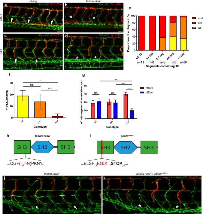
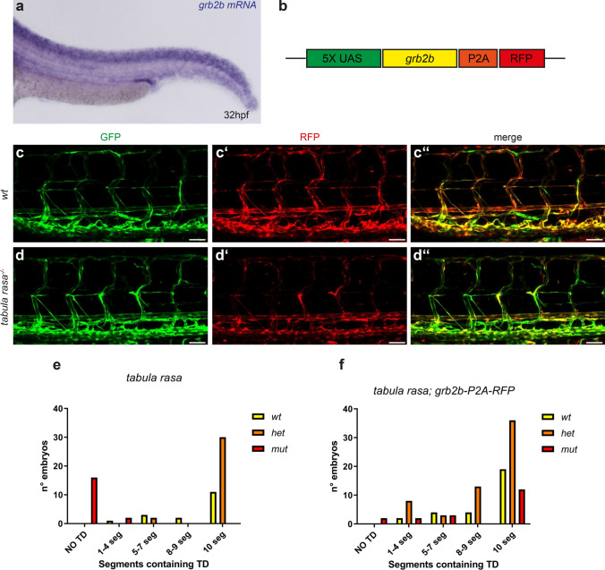
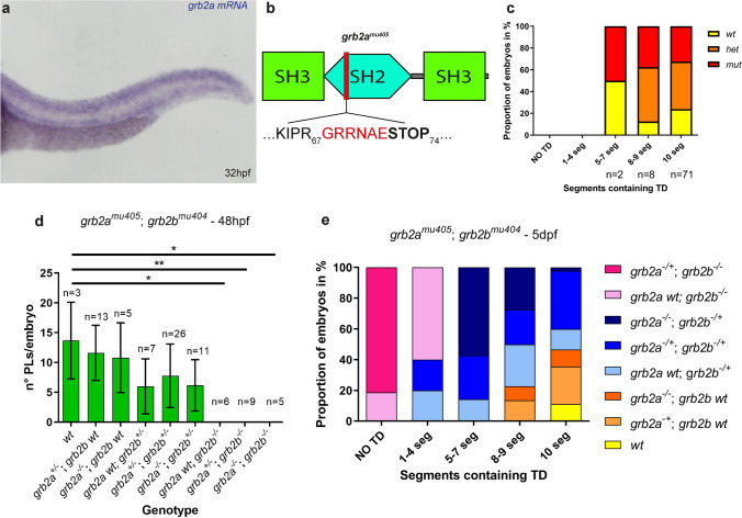
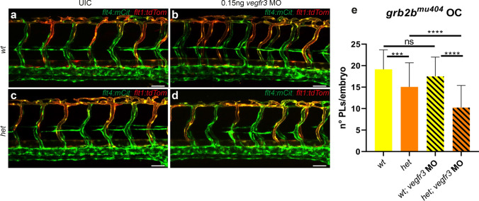
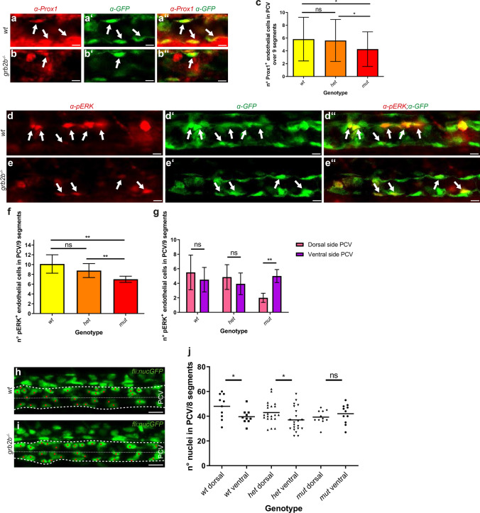
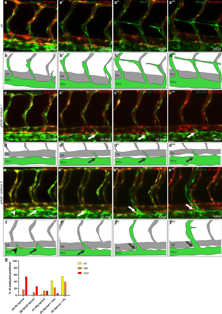
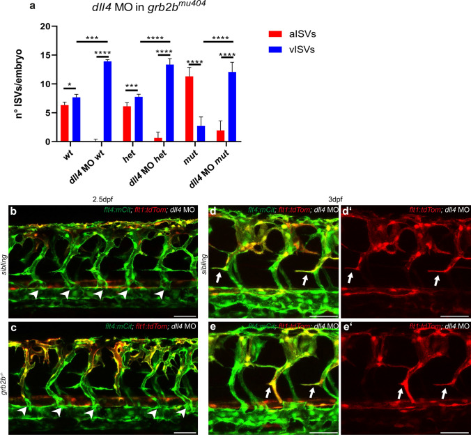
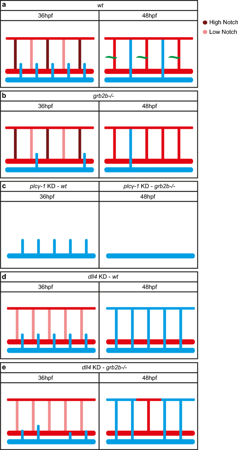
Vegfc/Vegfr3 signaling is critical for lymphangiogenesis, the sprouting of lymphatic vessels. In zebrafish, cells sprouting from the posterior cardinal vein can either form lymphatic precursor cells or contribute to intersegmental vein formation. Both, the Vegfc-dependent differential induction of Prox1a in sprouting cells as well as a Notch-mediated pre-pattern within intersegmental vessels have been associated with the regulation of secondary sprout behavior. However, how exactly a differential lymphatic versus venous sprout cell behavior is achieved is not fully understood. Here, we characterize a zebrafish mutant in the adaptor protein Grb2b, and demonstrate through genetic interaction studies that Grb2b acts within the Vegfr3 pathway. Mutant embryos exhibit phenotypes that are consistent with reduced Vegfr3 signaling outputs prior to the sprouting of endothelial cells from the vein. During secondary sprouting stages, loss of grb2b leads to defective cell behaviors resulting in a loss of parachordal lymphangioblasts, while only partially affecting the number of intersegmental veins. A second GRB2 zebrafish ortholog, grb2a, contributes to the development of lymphatic structures in the meninges and in the head, but not in the trunk. Our results illustrate an essential role of Grb2b in vivo for cell migration to the horizontal myoseptum and for the correct formation of the lymphatic vasculature, while being less critically required in intersegmental vein formation. Thus, there appear to be higher requirements for Grb2b and therefore Vegfr3 downstream signaling levels in lymphatic versus vein precursor-generating sprouts.

Keywords: Angiogenesis; Development; Lymphangiogenesis; VEGFR3; Vasculature; Zebrafish.


Conflict of interest statement

The authors declare no competing interests.

Figures

Fig. 1
Fig. 1
Mutations in the grb2b gene interfere with lymphatic development. ad Confocal projections of the trunk region in siblings (a, c) and tabula rasa mutants (b, d) at 48hpf (a, b) and at 5dpf (c, d) with flt4:mCitrine highlighting venous and lymphatic structures in green and the flt1:tdTomato transgene showing arterial endothelial cells in red. Arrows indicate the presence of PLs at the HM (a) and of a fully developed TD (c) in siblings, whereas asterisks indicate the lack of PLs (b) and of TD fragments (d) in mutants. e Quantification of TD-containing segments scored over the length of 10 consecutive somites at 5dpf. Most tabula rasa mutants lack the whole TD or they form only few fragments. wt: n = 24; het: n = 45; mut: n = 27. f Quantification of the number of PLs per embryo at 48hpf shows a significant decrease in the number of PLs in tabula rasa mutants compared to both wild types and heterozygotes. wt: n = 4; het: n = 12; mut: n = 6. ** Between wt and mut: P value 0.0095 (Mann–Whitney). ***Between het and mut: P value 0.003 (Mann–Whitney). g Quantification of the number of aISVs and vISVs in 20 consecutive segments (bilateral) at 48hpf in the zebrafish trunk. tabula rasa mutants display significantly higher numbers of arteries than veins. wt n = 4; het: n = 12; mut: n = 6. **Between vISVs wt and vISVs mut: P value = 0.0048 (Mann–Whitney). ***Between vISVs het and vISVs mut: P value  = 0.0001 (Mann Whitney). ** Between aISVs mut and vISVs mut: P value 0.0022 (Mann–Whitney). h, i Schematic representations of the Grb2b protein containing two SH3 domains and one SH2 domain. h I48 > N indicates the point mutation in the tabula rasa mutant: the isoleucine at position 48 is replaced by an asparagine. i A 2 bp deletion in the grb2bmu404 allele is predicted to cause a frame-shift after amino acid 19, within the first SH3 domain of the protein. j, k Confocal projections showing the lack of complementation in tabula rasa and grb2bmu404 trans-heterozygous embryos. j tabula rasa heterozygous embryos have a full TD, as indicated by arrows. k tabula rasa+/−; grb2bmu404+/− trans-heterozygous embryos lack the TD (indicated by asterisks). flt4:mCitrine is shown in green and flt1:tdTomato in red. SH2 Src homology domain 2, SH3 Src homology domain 3, aISVs arterial intersegmental vessels, vISVs venous intersegmental vessels, PL parachordal lymphangioblast, HM horizontal myoseptum, TD thoracic duct, ns not significant. Scale bars: 50 µm. Data in f, g are mean ± sd
Fig. 2
Fig. 2
Endothelial-specific expression of grb2b is sufficient for normal lymphatic development. a In situ hybridization of grb2b shows expression in the majority of tissues in zebrafish embryos at 32hpf. b Rescue construct containing a 5xUAS element upstream of the grb2b cDNA which was fused with an RFP cassette via a P2A self-cleaving peptide. cdʺ Confocal projections of transgenic flt4:Gal4; UAS:GFP; UAS:grb2b-P2A-RFP embryos from a tabula rasa in-cross at 5dpf. A tabula rasa wild-type embryo is shown in ccʺ and a homozygous mutant in ddʺ. e, f Quantification of trunk segments containing TD (scored over the length of 10 somites) in 5dpf embryos from a tabula rasa+/−; flt4:Gal4; UAS:GFP; grb2b-P2A-RFP in-cross. e GFP+ RFP embryos (i.e., not containing the rescue construct) served as a control. wt: n = 17; het: n = 32; mut: n = 18. f The mutant phenotype is rescued in embryos expressing the rescue construct (GFP+ and RFP+). wt: n = 29; het: n = 60; mut: n = 19. TD thoracic duct, wt wild type; het heterozygous; mut mutant for tabula rasa. Scale bars: 50 µm
Fig. 3
Fig. 3
Loss of grb2a does not cause lymphatic defects in the trunk, but Grb2a can functionally compensate for the loss of grb2b upon over-expression. a In situ hybridization of grb2a shows wide expression in the zebrafish embryo at 32hpf. b Schematic representation of the Grb2a protein, with two SH3 domains and one SH2 domain, highlighting the − 5 bp mutation in the SH2 domain of the grb2amu405 allele. c Quantification of TD fragments shows that grb2a is not essential for trunk lymphatic development. The presence of TD was quantified over 10 segments. wt: n = 19; het: n = 35; mut: n = 27. d Quantification of the number of PLs at 48hpf in the different genotypes of a grb2amu405; grb2bmu404 double heterozygous in-cross. Lymphatic precursor cells do not form when embryos are mutant for grb2b, independent of the number of grb2a wild-type copies. * Between wt and grb2a wt; grb2b−/−: P value 0.0119 (Mann–Whitney); **Between wt and grb2a+/−; grb2b−/−: P value = 0.0045 (Mann–Whitney); *Between wt and grb2a−/−; grb2b−/−: P value = 0.0179 (Mann–Whitney). Note that the comparison between wt and each one of the other genotypes resulted in a non-significant difference. e Quantification of TD+ segments in embryos from grb2amu405; grb2bmu404 double heterozygous parents at 5dpf. The development of the main lymphatic vessel depends on grb2b, and not on grb2a. d, e wt: n = 5; grb2a+/−; grb2b wt: n = 14; grb2a−/−; grb2b wt: n = 7; grb2a wt; grb2b+/−: n = 14; grb2a+/−; grb2b+/−: n = 25; grb2a−/−; grb2b+/−: n = 11; grb2a wt; grb2b−/−: n = 6; grb2a+/−; grb2b−/−: n = 13. SH2 Src homology domain 2, SH3 Src homology domain 3, PL parachordal lymphangioblast, TD thoracic duct. Data in d represent the mean ± sd
Fig. 4
Fig. 4
grb2b is a member of the Vegfc/Vegfr3 signaling pathway. ae Injections of 0.15 ng vegfr3 MO in embryos from a grb2bmu404 outcross. ad Confocal pictures of 48hpf old embryos; flt4:mCitrine is shown in green and flt1:tdTomato in red. e There is no difference in the number of PLs between un-injected wt embryos and wt embryos injected with 0.15 ng of vegfr3 MO. grb2bmu404 heterozygous embryos show a significant decrease in PLs after injection of 0.15 ng of vegfr3 MO. wt UIC: n = 45; wt 0.15 ng vegfr3 MO: n = 51; het UIC: n = 49; het 0.15 ng vegfr3 MO: n = 43. ***Between wt and het: P value = 0.0002 (t test, two tailed); ***Between wt 0.15 ng vegfr3 MO and het 0.15 ng vegfr3 MO: P value < 0.0001 (Mann–Whitney); ****Between het and het 0.15 ng vegfr3 MO: P value < 0.0001 (t-test, two tailed), ns not significant, UIC un-injected control, MO morpholino, OC outcross, PL parachordal lymphangioblast. Scale bars: 50 μm. Data in e are mean ± sd
Fig. 5
Fig. 5
grb2b mutants show a defect in PCV polarization at 32hpf and exhibit decreased numbers of Prox1 and pERK positive endothelial cells. ac The number of Prox1+ endothelial cells is significantly reduced in grb2bmu404 mutants at 32hpf. abʺ Confocal projections of the PCV at 32hpf in wild-type and mutant embryos. Prox1 antibody staining is shown in red and flt4:mCitrine in green, with arrows pointing at cells that co-express Prox1 and Flt4. c Quantification of Prox1+ endothelial cells in the PCV at 32hpf across 9 segments. wt: n = 38; het: n = 72; mut: n = 33. *Between wt and mut: P value = 0.0373 (t test, two-tailed); *Between het and mut: P value = 0.0412 (t test, two-tailed). df pERK+ endothelial cells are reduced in numbers in grb2bmu404 mutants at 32hpf. deʺ Confocal projections showing pERK antibody staining in red and endothelial cells in green (flt4:mCitrine) in the PCV at 32hpf. Arrows point at cells that express both pERK and Flt4. f Quantification of the number of pERK+ cells across 9 segments, demonstrating a decreased number in grb2b mutants. wt: n = 8; het: n = 14; mut: n = 6. **Between wt and mut: P value = 0.0013 (Mann–Whitney); ** between het and mut: P value 0.0036 (Mann–Whitney). g Moreover, pERK+ cells in wild type and heterozygotes are equally distributed between dorsal and ventral hemispheres of the PCV, whereas in mutants they are more concentrated in the ventral side. wt: n = 8; het: n = 14; mut: n = 6. **Between dorsal side and ventral side in mut: P value = 0.0022. h, i Confocal projections of fli:nEGFP wild-type and mutant embryos, highlighting the PCV between the more prominent dashed lines. The thinner dashed lines divide the PCV in two equal parts: the dorsal and the ventral side. j Quantifications of nuclei in the PCV at 32hpf showing that in grb2bmu404 mutants, endothelial cells are equally distributed between the ventral and the dorsal side. In siblings, endothelial cells are enriched in the dorsal part of the PCV. wt: n = 10; het: n = 21; mut: n = 11. *Between wt dorsal and wt ventral: P value 0.0327 (t test, two-tailed); *Between het dorsal and het ventral: P value  = 0.044 (t test, two-tailed). ns not significant. Scale bars: abʺ: 10 µm; deʺ: 10 µm; h, i 20 µm. Data in c, f, g are mean ± s.d. Data in j represent the mean with individual data points. All quantifications have been done blindly
Fig. 6
Fig. 6
Secondary sprouting is defective in grb2b mutant embryos. afʹʺ Still images taken from overnight videos (see Supplementary movies 1–3) and corresponding schematic cartoons of wild type (abʹʺ) and grb2bmu404 mutant (cfʹʺ) embryos between 30hpf and 48hpf, showing different sprout behaviors. a In wild-type embryos, cells sprout from the PCV and either form PLs at the HM or remodel an intersegmental artery into a vein. c, e Still images of two different grb2b mutant embryos. The arrow in cʹ–dʹʺ highlights a cell attempting to sprout from the PCV and extending towards an aISV, but failing to establish a stable connection. The arrowhead in e and f points at an endothelial cell with a small filopodium that retracts. The arrows in pictures from e to fʹʺ point at an endothelial sprout that reaches an aISV, establishes a stable connection and remodels the artery into a vein. In all images flt4:mCitrine is shown in green and flt1:tdTomato in red. g Quantification of the different sprout behaviors observed between 31 and 48hpf (indicated as percentage). For quantification, both sides of a four segments stretch per embryo were analyzed. wt: n = 5, het: n = 16, mut: n = 23. Scale bars: 25 µm. Data in d are mean ± sd
Fig. 7
Fig. 7
In the absence of arteries, cells fail to sprout in grb2b mutants. ac Confocal projections of plcγ-1 MO injected embryos. All vessels are shown in green (fli:GFP). d Sprout quantification at 48hpf of embryos from a tabula rasa in-cross, injected with a plcγ-1 MO. plcγ-1 MO injection inhibits primary sprouting, allowing quantification/visualization of secondary sprouts in absence of arteries. The number of lympho-venous sprouts is significantly decreased in tabula rasa mutants. wt: n = 4; het: n = 32; mut: n = 24. ****Between wt and mut: P value  < 0.0001 (Mann–Whitney); ****Between het and mut: P value < 0.0001 (Mann–Whitney); ns not significant. MO morpholino. Scale bars: 50 µm. Data in d are mean ± sd
Fig. 8
Fig. 8
grb2b mutants show an increase in vISVs upon dll4 knockdown. a Quantifications of aISVs and vISVS at 2.5dpf in un-injected control and dll4 MO injected embryos from grb2bmu404 heterozygous parents. 14 ISVs per embryo were analyzed. *Between aISVs and vISV in wt: P value 0.013 (Mann–Whitney); ****Between aISVs and vISV in dll4 MO wt: P value < 0.0001 (Mann–Whitney); ***Between aISVs and vISV in het: P value 0.0009 (Mann–Whitney); ****Between aISVs and vISV in dll4 MO het: P value < 0.0001 (Mann–Whitney); ****Between aISVs and vISV in mut: P value < 0.0001 (Mann–Whitney); ****Between aISVs and vISV in dll4 MO mut: P value < 0.0001 (Mann–Whitney); ***Between vISVs wt and vISVs dll4 MO wt: P value = 0.0001 (Mann–Whitney); ****Between vISVs het and vISV in dll4 MO het: P value < 0.0001 (Mann–Whitney); ****Between vISVs mut and vISV in dll4 MO mut: P value < 0.0001 (Mann–Whitney). beʹ Confocal pictures of grb2b mutant or sibling embryos at 2.5dpf (b, c) and 3dpf (deʹ), injected with dll4 MO. Veins are shown in green (flt4:mCitrine) and arteries in red (flt1:tdTomato). Arrowheads highlight vISVs while arrows mark protrusions from ISVs extending towards the HM. aISV arterial intersegmental vessel, vISV venous intersegmental vessel, MO morpholino. Scale bars: 50 µm. Data in a represent mean ± sd
Fig. 9
Fig. 9
grb2b is essential for cells to sprout from the PCV and for PL formation, but not for vein formation. a In wild-type embryos, secondary sprouts are either forming PLs, shown in green (if they are close to an aISV with high Notch signaling levels), or a vISV (in case the aISV has low levels of Notch activity). b grb2b mutant embryos show defective secondary sprouting. If a cell protruding from the PCV encounters an intersegmental artery with high Notch levels, then it will retract towards the PCV, not being able to migrate to the HM. If the cell makes contact with a low Notch signaling artery, it will form an intersegmental vein. c Upon plcγ-1 knockdown, no intersegmental arteries develop. In wild-type embryos sprouts normally migrate out from the PCV, whereas no stable sprout is detected at 48hpf in grb2b mutants. d, e Upon dll4 knockdown most of aISVs are remodeled into vISVs in both wild-type and grb2b mutant embryos, indicating that a connection between a cell in the PCV and the intersegmental artery is sufficient for a venous endothelial cell to form a vein

Similar articles

Cited by

References

    1. Mauri C, Wang G, Schulte-Merker S. From fish embryos to human patients: lymphangiogenesis in development and disease. Curr Opin Immunol. 2018;53:167–172. doi: 10.1016/j.coi.2018.05.003. - DOI - PubMed
    1. Alitalo K. The lymphatic vasculature in disease. Nat Med. 2011;17(11):1371–1380. doi: 10.1038/nm.2545. - DOI - PubMed
    1. Joukov V, Sorsa T, Kumar V, Jeltsch M, Claesson-Welsh L, Cao Y, Saksela O, Kalkkinen N, Alitalo K. Proteolytic processing regulates receptor specificity and activity of VEGF-C. EMBO J. 1997;16(13):3898–3911. doi: 10.1093/emboj/16.13.3898. - DOI - PMC - PubMed
    1. Le Guen L, Karpanen T, Schulte D, Harris NC, Koltowska K, Roukens G, Bower NI, van Impel A, Stacker SA, Achen MG, Schulte-Merker S, Hogan BM. Ccbe1 regulates Vegfc-mediated induction of Vegfr3 signaling during embryonic lymphangiogenesis. Development. 2014;141(6):1239–1249. doi: 10.1242/dev.100495. - DOI - PubMed
    1. Jeltsch M, Jha SK, Tvorogov D, Anisimov A, Leppanen VM, Holopainen T, Kivela R, Ortega S, Karpanen T, Alitalo K. CCBE1 enhances lymphangiogenesis via A disintegrin and metalloprotease with thrombospondin motifs-3-mediated vascular endothelial growth factor-C activation. Circulation. 2014;129(19):1962–1971. doi: 10.1161/CIRCULATIONAHA.113.002779. - DOI - PubMed

Publication types

MeSH terms

Substances

LinkOut - more resources