. 2021 Jul;36(7):2510-2517.e6.
doi: 10.1016/j.arth.2021.02.026. Epub 2021 Feb 16.

A Deep Learning Tool for Automated Radiographic Measurement of Acetabular Component Inclination and Version After Total Hip Arthroplasty

Affiliations

A Deep Learning Tool for Automated Radiographic Measurement of Acetabular Component Inclination and Version After Total Hip Arthroplasty

Pouria Rouzrokh et al. J Arthroplasty. 2021 Jul.

Abstract

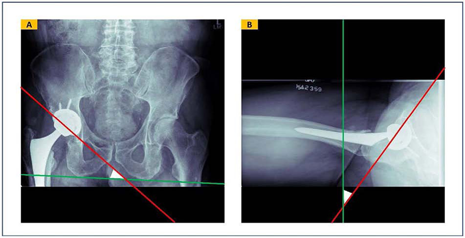
Background: Inappropriate acetabular component angular position is believed to increase the risk of hip dislocation after total hip arthroplasty. However, manual measurement of these angles is time consuming and prone to interobserver variability. The purpose of this study was to develop a deep learning tool to automate the measurement of acetabular component angles on postoperative radiographs.

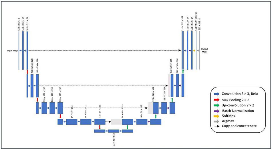
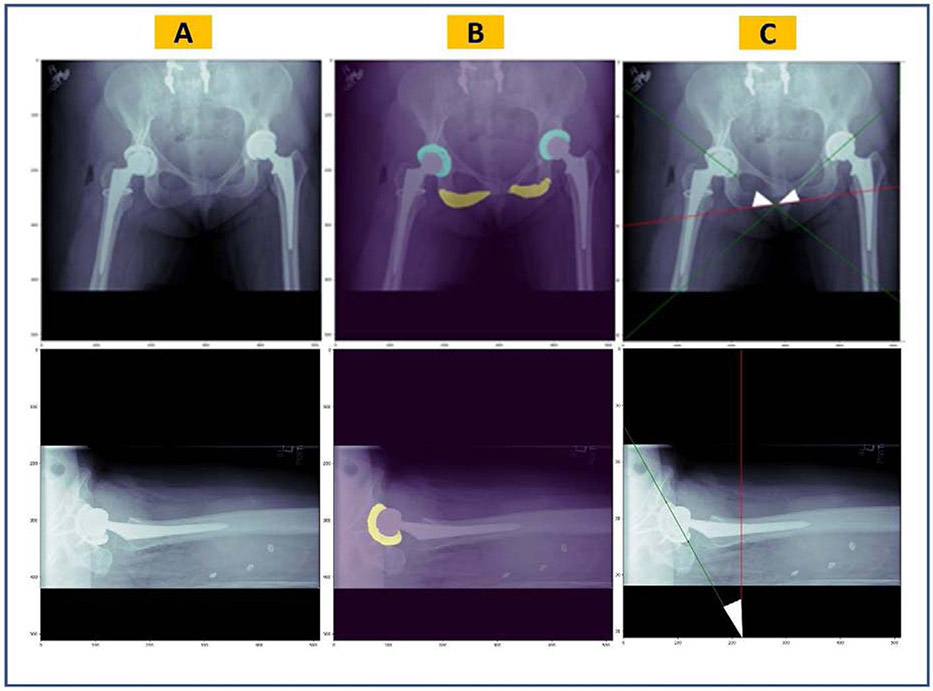
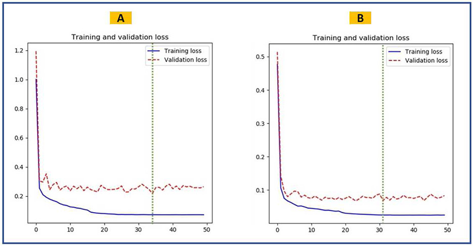
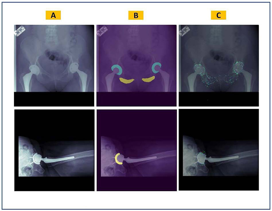
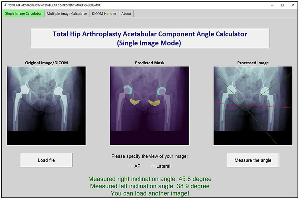
Methods: Two cohorts of 600 anteroposterior (AP) pelvis and 600 cross-table lateral hip postoperative radiographs were used to develop deep learning models to segment the acetabular component and the ischial tuberosities. Cohorts were manually annotated, augmented, and randomly split to train-validation-test data sets on an 8:1:1 basis. Two U-Net convolutional neural network models (one for AP and one for cross-table lateral radiographs) were trained for 50 epochs. Image processing was then deployed to measure the acetabular component angles on the predicted masks for anatomical landmarks. Performance of the tool was tested on 80 AP and 80 cross-table lateral radiographs.

Results: The convolutional neural network models achieved a mean Dice similarity coefficient of 0.878 and 0.903 on AP and cross-table lateral test data sets, respectively. The mean difference between human-level and machine-level measurements was 1.35° (σ = 1.07°) and 1.39° (σ = 1.27°) for the inclination and anteversion angles, respectively. Differences of 5⁰ or more between human-level and machine-level measurements were observed in less than 2.5% of cases.

Conclusion: We developed a highly accurate deep learning tool to automate the measurement of angular position of acetabular components for use in both clinical and research settings.

Level of evidence: III.

Keywords: acetabular component angle; anteversion angle; artificial intelligence; deep learning; inclination angle; total hip arthroplasty.


Figures

FIGURE 1.
FIGURE 1.
Inclination and anteversion acetabular component angles defined in a radiology reference system. (A) Inclination angle is defined as the angle between the acetabular component longitudinal axis and the trans-ischial tuberosity line on an anteroposterior radiograph. (B) Anteversion angle is defined by the angle between the acetabular component longitudinal axis and a standard vertical line perpendicularly drawn to the table on a hip cross-table lateral radiograph.
Figure 2.
Figure 2.
Architecture of U-Net CNN model used to segment the radiographic images. Encoder of the model had the VGG-16 architecture and its initial weights were pooled from a model pre-trained on the ImageNet database. The output of the model will initially have three channels (in AP model) or two channels (in cross-table lateral model). An argmax function will change this output to a one-channel 512×512-pixel mask, that is then used for image processing.
Figure 3.
Figure 3.
Overview of the pipeline for automatic measurement of the acetabular component angles. (A) Original radiographic images, (B) predicted masks by the semantic segmentation U-Net models overlaid on the original images, (C) acetabular component longitudinal axes (in green) and the trans-ischial tuberosity line or standard vertical line (in red) which are estimated by image processing. Together, they form the inclination angle on AP pelvis images and anteversion angle on hip cross-table lateral images (white triangles).
Figure 4.
Figure 4.
Training performance of the semantic segmentation U-Net models. The green dashed line shows the epoch when the best model was saved. (A) Training and validation loss curves for the inclination model. (B) Training and validation loss curves for the anteversion model.
Figure 5.
Figure 5.
Visualization of the semantic segmentation U-Net models overlaid on sample original images. (A) Original radiographic images, (B) predicted masks, (C) integrated gradients maps where the red color highlights the most influential pixels on the model’s predictions.
Figure 6.
Figure 6.
Screenshot from the Total Hip Arthroplasty Acetabular Component Angle Calculator software, a tool developed to deploy the semantic segmentation U-Net models and their subsequent image processing workflow into a stand-alone graphic user interface (GUI). The software can measure acetabular component angles on single or multiple PNG image (or DICOM) files.

References

    1. Learmonth ID, Young C, Rorabeck C. The operation of the century: total hip replacement. Lancet 2007;370:1508–19. 10.1016/S0140-6736(07)60457-7. - DOI - PubMed
    1. Bozic KJ, Kurtz SM, Lau E, Ong K, Vail TP, Berry DJ. The epidemiology of revision total hip arthroplasty in the United States. J Bone Joint Surg Am 2009;91:128–33. 10.2106/JBJS.H.00155. - DOI - PubMed
    1. Kunutsor SK, Barrett MC, Beswick AD, Judge A, Blom AW, Wylde V, et al. Risk factors for dislocation after primary total hip replacement: a systematic review and meta-analysis of 125 studies involving approximately five million hip replacements. Lancet Rheumatol 2019;1:e111–21. 10.1016/S2665-9913(19)30045-1. - DOI - PubMed
    1. Abdel MP, Cross MB, Yasen AT, Haddad FS. The functional and financial impact of isolated and recurrent dislocation after total hip arthroplasty. Bone Joint J 2015;97-B:1046–9. 10.1302/0301-620X.97B8.34952. - DOI - PubMed
    1. Biedermann R, Tonin A, Krismer M, Rachbauer F, Eibl G, Stöckl B. Reducing the risk of dislocation after total hip arthroplasty: the effect of orientation of the acetabular component. J Bone Joint Surg Br 2005;87:762–9. 10.1302/0301-620X.87B6.14745. - DOI - PubMed

Publication types