

Calcific tendonitis of the flexor pollicis longus tendon at the thumb interphalangeal joint in childhood
- PMID: 33678974
- PMCID: PMC7901431
- DOI: 10.1080/08998280.2020.1834805
Calcific tendonitis of the flexor pollicis longus tendon at the thumb interphalangeal joint in childhood
Abstract
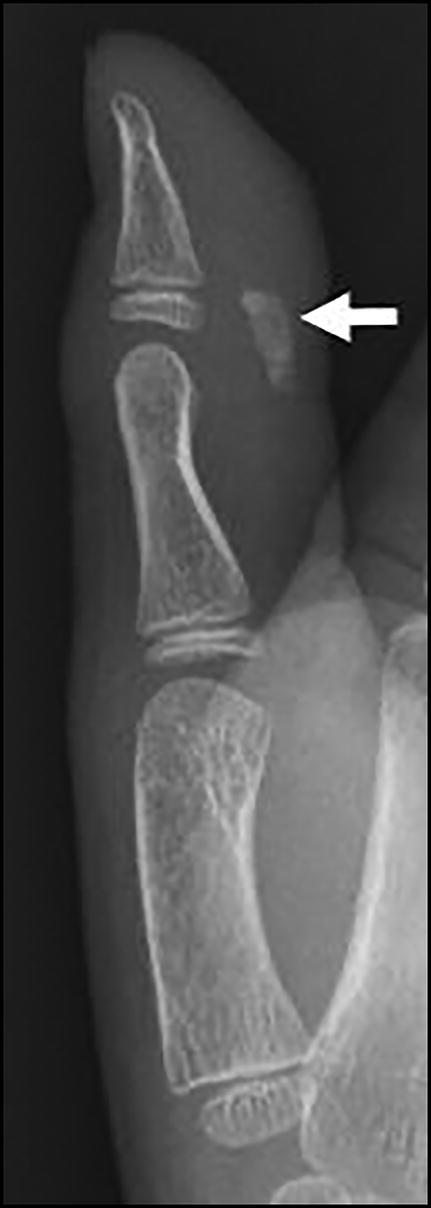
In rare instances, calcific tendonitis may manifest in the pediatric population as inflammatory calcium hydroxyapatite deposition. To our knowledge, there have been no previous case reports involving the flexor pollicis longus tendon at the thumb interphalangeal joint. We present a 9-year-old boy with a painful mass at the right thumb interphalangeal joint. Initial radiographs revealed a 7-mm ovoid calcific mass along the volar soft tissues of the thumb interphalangeal joint. Subsequent ultrasound and magnetic resonance findings further confirmed calcification with surrounding edema. Because the pain was limiting the patient's school activities, his family elected for excisional biopsy of the calcific mass. Pathology ultimately revealed prominent dystrophic calcifications with surrounding granulomatous inflammation, consistent with calcific tendonitis.
Keywords: Calcific tendonitis; flexor pollicis tendon; magnetic resonance imaging; pediatric; radiograph; ultrasound.
Copyright © 2020 Baylor University Medical Center.
Figures


Similar articles
-
Flexor pollicis longus dysfunction after volar plate fixation of distal radius fractures.J Hand Surg Am. 2013 Sep;38(9):1691-7. doi: 10.1016/j.jhsa.2013.06.005. Epub 2013 Jul 30. J Hand Surg Am. 2013. PMID: 23910382
-
Irreducible dislocation of the thumb interphalangeal joint due to displaced flexor pollicis longus tendon: case report and new reduction technique.Arch Orthop Trauma Surg. 2014 Aug;134(8):1175-8. doi: 10.1007/s00402-014-2024-6. Epub 2014 Jun 6. Arch Orthop Trauma Surg. 2014. PMID: 24902518
-
Radiologic findings of the flexor pollicis longus hypoplasia.Skeletal Radiol. 2007 Jun;36 Suppl 1:120-3. doi: 10.1007/s00256-006-0187-0. Epub 2006 Oct 7. Skeletal Radiol. 2007. PMID: 17028901
-
Complex Irreducible Dorsal Dislocation of Thumb Interphalangeal Joint: Controversies and Consensus in Management.J Hand Surg Asian Pac Vol. 2020 Sep;25(3):378-383. doi: 10.1142/S2424835520720133. J Hand Surg Asian Pac Vol. 2020. PMID: 32723059 Review.
-
Reconstruction of the rheumatoid thumb.Hand Clin. 1992 Feb;8(1):121-9. Hand Clin. 1992. PMID: 1572917 Review.
References
Publication types
LinkOut - more resources
Full Text Sources