

Pericardial agenesis
- PMID: 33679077
- PMCID: PMC7918029
- DOI: 10.4103/apc.APC_152_19
Pericardial agenesis
Abstract
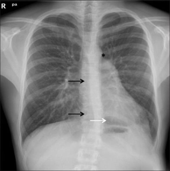
Congenital pericardial defect is a rare and usually asymptomatic condition which is classified incomplete or partial. Up to 70% of cases consist of complete absence of left pericardium. The diagnosis may be challenging due to its low frequency and absence of correlation with any specific finding on the clinical examination. Cardiac magnetic resonance imaging is the gold standard imaging technique for the diagnosis confirming the absence of pericardium, although other indirect signs may be seen. In partial defects, surgery is the treatment option. We present an incidental finding of total agenesis of the left pericardium in an asymptomatic 16-year-old male diagnosed in a preoperative assessment of a bone fracture.
Keywords: Magnetic resonance imaging; pediatrics; pericardial disease.
Copyright: © 2020 Annals of Pediatric Cardiology.
Conflict of interest statement
There are no conflicts of interest.
Figures


References
-
- Cuccuini M, Lisi F, Consoli A, Mancini S, Bellino V, Galanti G, et al. Congenital defects of pericardium: Case reports and review of literature. Ital J Anat Embryol. 2013;118:136–50. - PubMed
-
- Shah AB, Kronzon I. Congenital defects of the pericardium: A review. Eur Heart J Cardiovasc Imaging. 2015;16:821–7. - PubMed
Publication types
LinkOut - more resources
Full Text Sources
Other Literature Sources