

Effects of Recombinant Angiogenin on Collagen Fiber Formation and Angiogenesis in the Dermis of Wistar Rats
- PMID: 33679135
- PMCID: PMC7926187
- DOI: 10.2147/CCID.S294825
Effects of Recombinant Angiogenin on Collagen Fiber Formation and Angiogenesis in the Dermis of Wistar Rats
Abstract
Purpose: The purpose of this study was to assess the capability of recombinant angiogenin isolated from Pichia pastoris yeasts to stimulate regenerative processes in the dermis of experimental animals.
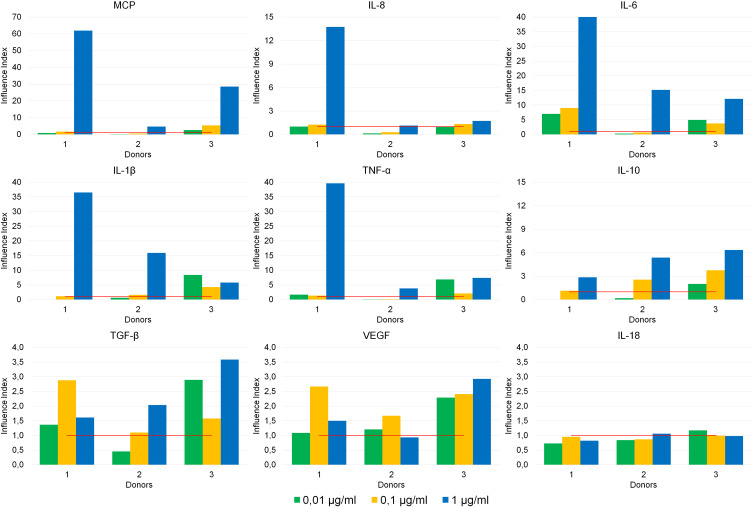
Patients and methods: Wistar rats were administered with recombinant angiogenin intracutaneously. Morphological examination of the skin and the assessment of the proliferative activity of the epidermal cells were carried out. Additionally, cytokine production by human whole blood cells exposed to angiogenin was analyzed ex vivo.
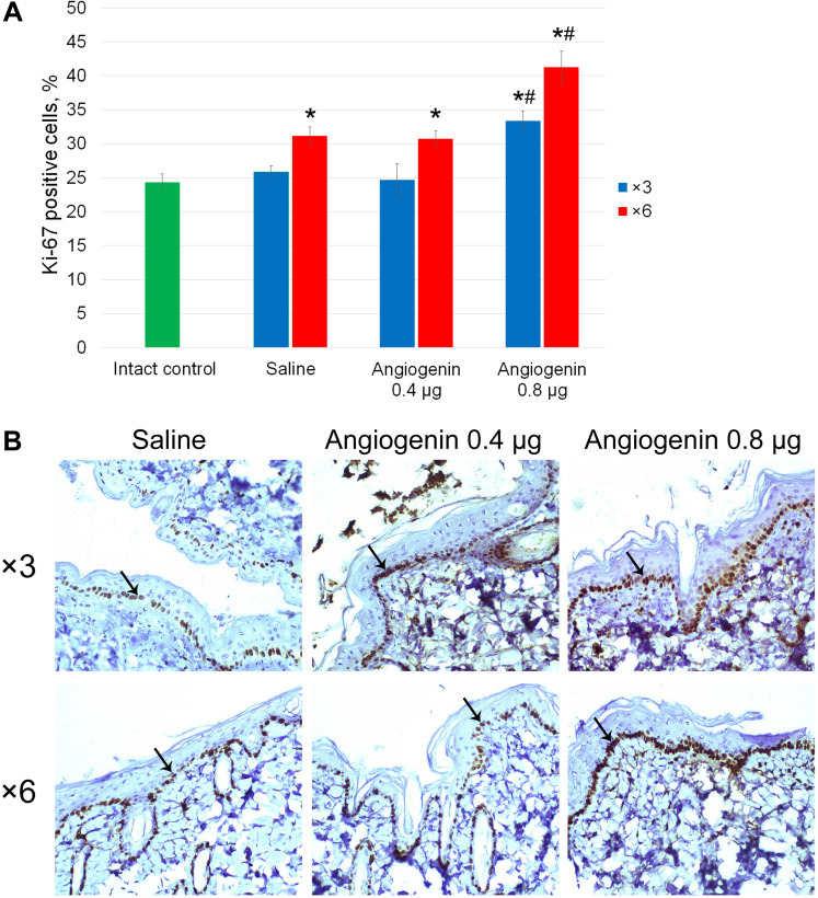
Results: Administration of angiogenin stimulates collagen fiber formation and angiogenesis. This stimulation is tightly associated with an increase in the number of fibroblasts, an increased numerical density of dermal blood vessels and an increased density of collagen fibers; also, it activates the proliferation of basal cells. Angiogenin induces the production of MCP, IL-8, IL-6, IL-1β, TNF-α, IL-10, TGF-β, and VEGF by blood cells.
Conclusion: The results obtained indicate a broad spectrum of actions of recombinant angiogenin during regenerative processes in the basal layer of the dermis.
Keywords: cytokines; intracutaneous injections; pathomorphological analysis; regeneration.
© 2021 Yurina et al.
Conflict of interest statement
The authors report no conflicts of interest in this work.
Figures

 Expansion of the stratum corneum,
  an increase in the number of epidermal layers,
  thickened collagen fibers,
  the site of newly formed collagen with proliferating fibroblasts (the edge of the injection area). Hematoxylin/eosin staining, 100× magnification.
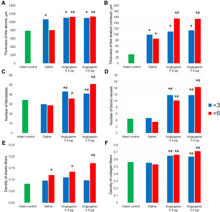
Similar articles
-
Angiogenin is expressed in human dermal papilla cells and stimulates hair growth.Arch Dermatol Res. 2009 Feb;301(2):139-49. doi: 10.1007/s00403-008-0907-5. Epub 2008 Oct 21. Arch Dermatol Res. 2009. PMID: 18936943
-
Angiogenin ameliorates corneal opacity and neovascularization via regulating immune response in corneal fibroblasts.BMC Ophthalmol. 2016 May 17;16:57. doi: 10.1186/s12886-016-0235-z. BMC Ophthalmol. 2016. PMID: 27356868 Free PMC article.
-
[Effect of a gel based on recombinant human angiogenin on the healing of donor palate wounds].Stomatologiia (Mosk). 2019;98(1):34-37. doi: 10.17116/stomat20199801134. Stomatologiia (Mosk). 2019. PMID: 30830091 Russian.
-
Targeting tumor micro-environment for design and development of novel anti-angiogenic agents arresting tumor growth.Prog Biophys Mol Biol. 2013 Nov;113(2):333-54. doi: 10.1016/j.pbiomolbio.2013.10.001. Epub 2013 Oct 15. Prog Biophys Mol Biol. 2013. PMID: 24139944 Review.
-
[Angiogenin and its role in angiogenesis].Mol Biol (Mosk). 2001 May-Jun;35(3):349-71. Mol Biol (Mosk). 2001. PMID: 11443914 Review. Russian.
Cited by
-
Angiogenin as a Possible Mediator of Macrophage-Mediated Regulation of Fibroblast Functions.Bull Exp Biol Med. 2023 Sep;175(5):658-661. doi: 10.1007/s10517-023-05921-z. Epub 2023 Oct 20. Bull Exp Biol Med. 2023. PMID: 37861896
-
Vascularized characteristics and functional regeneration of three-dimensional cell reconstruction of oral mucosa equivalents based on vascular homeostasis phenotypic modification.J Tissue Eng. 2024 Sep 18;15:20417314241268912. doi: 10.1177/20417314241268912. eCollection 2024 Jan-Dec. J Tissue Eng. 2024. PMID: 39301507 Free PMC article.
-
Protective Effects of Recombinant Human Angiogenin in Keratinocytes: New Insights on Oxidative Stress Response Mediated by RNases.Int J Mol Sci. 2022 Aug 7;23(15):8781. doi: 10.3390/ijms23158781. Int J Mol Sci. 2022. PMID: 35955913 Free PMC article.
-
Angiogenin and Copper Crossing in Wound Healing.Int J Mol Sci. 2021 Oct 2;22(19):10704. doi: 10.3390/ijms221910704. Int J Mol Sci. 2021. PMID: 34639045 Free PMC article. Review.
-
Human Keratinocytes and Fibroblasts Co-Cultured on Silk Fibroin Scaffolds Exosomally Overrelease Angiogenic and Growth Factors.Cells. 2023 Jul 11;12(14):1827. doi: 10.3390/cells12141827. Cells. 2023. PMID: 37508492 Free PMC article.
References
LinkOut - more resources
Full Text Sources
Other Literature Sources
Miscellaneous
