Repetitive Peripheral Magnetic Stimulation of Wrist Extensors Enhances Cortical Excitability and Motor Performance in Healthy Individuals
- PMID: 33679314
- PMCID: PMC7930341
- DOI: 10.3389/fnins.2021.632716
Repetitive Peripheral Magnetic Stimulation of Wrist Extensors Enhances Cortical Excitability and Motor Performance in Healthy Individuals
Abstract
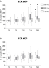
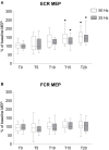
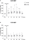
Repetitive peripheral magnetic stimulation (rPMS) may improve motor function following central nervous system lesions, but the optimal parameters of rPMS to induce neural plasticity and mechanisms underlying its action remain unclear. We examined the effects of rPMS over wrist extensor muscles on neural plasticity and motor performance in 26 healthy volunteers. In separate experiments, the effects of rPMS on motor evoked potentials (MEPs), short-interval intracortical inhibition (SICI), intracortical facilitation (ICF), direct motor response (M-wave), Hoffmann-reflex, and ballistic wrist extension movements were assessed before and after rPMS. First, to examine the effects of stimulus frequency, rPMS was applied at 50, 25, and 10 Hz by setting a fixed total number of stimuli. A significant increase in MEPs of wrist extensors was observed following 50 and 25 Hz rPMS, but not 10 Hz rPMS. Next, we examined the time required to induce plasticity by increasing the number of stimuli, and found that at least 15 min of 50 and 25 Hz rPMS was required. Based on these parameters, lasting effects were evaluated following 15 min of 50 or 25 Hz rPMS. A significant increase in MEP was observed up to 60 min following 50 and 25 Hz rPMS; similarly, an attenuation of SICI and enhancement of ICF were also observed. The maximal M-wave and Hoffmann-reflex did not change, suggesting that the increase in MEP was due to plastic changes at the motor cortex. This was accompanied by increasing force and electromyograms during wrist ballistic extension movements following 50 and 25 Hz rPMS. These findings suggest that 15 min of rPMS with 25 Hz or more induces an increase in cortical excitability of the relevant area rather than altering the excitability of spinal circuits, and has the potential to improve motor output.
Keywords: corticospinal tract; intracortical circuits; plasticity; rehabilitation; spinal networks; upper extremity.
Copyright © 2021 Nito, Katagiri, Yoshida, Koseki, Kudo, Nanba, Tanabe and Yamaguchi.
Conflict of interest statement
The authors declare that the research was conducted in the absence of any commercial or financial relationships that could be construed as a potential conflict of interest.
Figures


Similar articles
-
Effects of Repetitive Peripheral Magnetic Stimulation through Hand Splint Materials on Induced Movement and Corticospinal Excitability in Healthy Participants.Brain Sci. 2022 Feb 17;12(2):280. doi: 10.3390/brainsci12020280. Brain Sci. 2022. PMID: 35204043 Free PMC article.
-
Modulation of motor cortex excitability by paired peripheral and transcranial magnetic stimulation.Clin Neurophysiol. 2017 Oct;128(10):2043-2047. doi: 10.1016/j.clinph.2017.06.041. Epub 2017 Jul 17. Clin Neurophysiol. 2017. PMID: 28858700
-
Time course changes in corticospinal excitability during repetitive peripheral magnetic stimulation combined with motor imagery.Neurosci Lett. 2022 Feb 6;771:136427. doi: 10.1016/j.neulet.2021.136427. Epub 2021 Dec 28. Neurosci Lett. 2022. PMID: 34971770
-
Modulation of sensorimotor cortex by repetitive peripheral magnetic stimulation.Front Hum Neurosci. 2015 Jul 14;9:407. doi: 10.3389/fnhum.2015.00407. eCollection 2015. Front Hum Neurosci. 2015. PMID: 26236220 Free PMC article.
-
Effects of repetitive peripheral magnetic stimulation on normal or impaired motor control. A review.Neurophysiol Clin. 2013 Oct;43(4):251-60. doi: 10.1016/j.neucli.2013.05.003. Epub 2013 Jun 10. Neurophysiol Clin. 2013. PMID: 24094911 Review.
Cited by
-
Effects of repetitive peripheral magnetic stimulation vs. conventional therapy in the management of carpal tunnel syndrome: a pilot randomized controlled trial.PeerJ. 2023 May 18;11:e15398. doi: 10.7717/peerj.15398. eCollection 2023. PeerJ. 2023. PMID: 37220528 Free PMC article. Clinical Trial.
-
Effects of Different Intensities of Repetitive Peripheral Magnetic Stimulation on Spinal Reciprocal Inhibition in Healthy Persons.Juntendo Iji Zasshi. 2024 Jun 15;70(4):283-288. doi: 10.14789/jmj.JMJ23-0039-OA. eCollection 2024. Juntendo Iji Zasshi. 2024. PMID: 39431178 Free PMC article.
-
Effects of Repetitive Peripheral Magnetic Stimulation through Hand Splint Materials on Induced Movement and Corticospinal Excitability in Healthy Participants.Brain Sci. 2022 Feb 17;12(2):280. doi: 10.3390/brainsci12020280. Brain Sci. 2022. PMID: 35204043 Free PMC article.
-
Cortical Mechanisms Underlying Effects of Repetitive Peripheral Magnetic Stimulation on Dynamic and Static Postural Control in Patients with Chronic Non-Specific Low Back Pain: A Double-Blind Randomized Clinical Trial.Pain Ther. 2024 Aug;13(4):953-970. doi: 10.1007/s40122-024-00613-6. Epub 2024 Jun 19. Pain Ther. 2024. PMID: 38896200 Free PMC article.
-
Effects of repetitive peripheral magnetic stimulation for the upper limb after stroke: Meta-analysis of randomized controlled trials.Heliyon. 2023 Apr 22;9(5):e15767. doi: 10.1016/j.heliyon.2023.e15767. eCollection 2023 May. Heliyon. 2023. PMID: 37180919 Free PMC article. Review.
References
-
- Andrews R. K., Schabrun S. M., Ridding M. C., Galea M. P., Hodges P. W., Chipchase L. S. (2013). The effect of electrical stimulation on corticospinal excitability is dependent on application duration: a same subject pre-post test design. J. Neuroeng. Rehabil. 10:51. 10.1186/1743-0003-10-51 - DOI - PMC - PubMed
-
- Chapman L. J., Chapman J. P. (1987). The measurement of handedness. Brain Cogn. 6 175–183. - PubMed
LinkOut - more resources
Full Text Sources
Other Literature Sources

