. 2021 Feb 18:15:642521.
doi: 10.3389/fncel.2021.642521. eCollection 2021.

Low-Density Lipoprotein Receptor-Related Protein 1 (LRP1) as a Novel Regulator of Early Astroglial Differentiation

Affiliations

Low-Density Lipoprotein Receptor-Related Protein 1 (LRP1) as a Novel Regulator of Early Astroglial Differentiation

Ramona Romeo et al. Front Cell Neurosci. .

Abstract

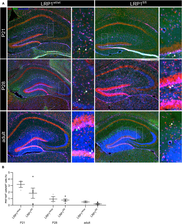
Astrocytes are the most abundant cell type within the central nervous system (CNS) with various functions. Furthermore, astrocytes show a regional and developmental heterogeneity traceable with specific markers. In this study, the influence of the low-density lipoprotein receptor-related protein 1 (LRP1) on astrocytic maturation within the hippocampus was analyzed during development. Previous studies mostly focused on the involvement of LRP1 in the neuronal compartment, where the deletion caused hyperactivity and motor dysfunctions in knockout animals. However, the influence of LRP1 on glia cells is less intensively investigated. Therefore, we used a newly generated mouse model, where LRP1 is specifically deleted from GLAST-positive astrocytes co-localized with the expression of the reporter tdTomato to visualize recombination and knockout events in vivo. The influence of LRP1 on the maturation of hippocampal astrocytes was assessed with immunohistochemical stainings against stage-specific markers as well as on mRNA level with RT-PCR analysis. The examination revealed that the knockout induction caused a significantly decreased number of mature astrocytes at an early developmental timepoint compared to control animals. Additionally, the delayed maturation of astrocytes also caused a reduced activity of neurons within the hippocampus. As previous studies showed that the glial specification and maturation of astrocytes is dependent on the signaling cascades Ras/Raf/MEK/Erk and PI3K/Akt, the phosphorylation of the signaling molecules Erk1/2 and Akt was analyzed. The hippocampal tissue of LRP1-deficient animals at P21 showed a significantly decreased amount of activated Erk in comparison to control tissue leading to the conclusion that the activation of this signaling cascade is dependent on LRP1 in astrocytes, which in turn is necessary for proper maturation of astrocytes. Our results showed that the deletion of LRP1 at an early developmental timepoint caused a delayed maturation of astrocytes in the hippocampus based on an altered activation of the Ras/Raf/MEK/Erk signaling pathway. However, with ongoing development these effects were compensated and the number of mature astrocytes was comparable as well as the activity of neurons. Therefore, LRP1 acts as an early regulator of the differentiation and maturation of astrocytes within the hippocampus.

Keywords: LRP1; astrocyte functions; astrocyte heterogeneity; differentiation; hippocampus; in vivo knockout model.


Conflict of interest statement

The authors declare that the research was conducted in the absence of any commercial or financial relationships that could be construed as a potential conflict of interest.

Figures

FIGURE 1
FIGURE 1
Knockout induction via lactating mothers was successful in vivo. The immunohistochemical staining against LRP1 (green) and tdTomato (red) revealed the expression of LRP1 by recombined astrocytes during development indicated by the arrowhead (A). The recombination rate differed between 85 and 97% during development in LRP1-deficient and control hippocampi (B). The knockout efficiency was evaluated by the quantification of LRP1- and tdTomato-double positive cells divided by the total number of recombined cells within the hippocampus. Here the quantification showed that the number of LRP1-expressing recombined cells was significantly decreased in the knockout compared to the control (C). Furthermore, the knockout induction was evaluated via RT-PCR (D). The gene expression of Lrp1 was not altered in LRP1-deficient hippocampi during development compared to control tissue (E). As a last approach the protein expression of LRP1 was analyzed with western blot analysis (F). The protein expression was comparable in both conditions during development (G) (Scale bar is 50 μm; mean ± SEM; two-way ANOVA with post-hoc Bonferroni test; *p < 0.05, **p < 0.01, ***p < 0.001; for N-values see Table 2).
FIGURE 2
FIGURE 2
Characterization of the knockout in vivo. To confirm that the deletion of LRP1 was restricted to the astrocytic lineage, an immunohistochemical staining against neurons (NeuN; green) and oligodendrocytes (CC1; green) was performed (A). There was no colocalization of both cell-specific markers with the expression of tdTomato in recombined cells indicated by the arrowhead leading to the assumption that the deletion of LRP1 only occurred in astrocytes. To further characterize the newly generated mouse line, the weights of the TAM-receiving animals were compared (B). The statistical analysis revealed no changes in the weight of knockout animals compared to the control. As the deletion of one member of the LDL-family might results in an increased expression of other members, the expression of Lrp2 (C,D) was investigated. However, the knockout induction of LRP1 caused no altered expression of LRP2 (Scale bar is 50 μm; mean ± SEM; two-way ANOVA with post-hoc Bonferroni test; for N-values see Table 2).
FIGURE 3
FIGURE 3
Proliferation capacity of LRP1-deficient astrocytes was not affected. The proliferation rate was evaluated with an immunohistochemical staining against phospho-Histone H3 (PH3; green) and tdTomato (A, red). Double-positive cells indicated by the arrowhead were quantified and the analysis showed that the numbers of proliferation events were comparable in both conditions (B, Scale bar is 50 μm; mean ± SEM; two-way ANOVA with post-hoc Bonferroni test; for N-values see Table 2).
FIGURE 4
FIGURE 4
Expression of late precursor or immature astrocytic genes was not altered in LRP1-deficient hippocampi. To investigate the late precursor stage, Glast (A,B) and Fgfr-3 (A,C) were analyzed, whereas the immature astrocytic stage was examined with the markers Aqp-4 (A,D) and Aldh1l1 (A,E). The analysis revealed no significant changes in the hippocampal tissue of LRP1-deficient animals compared to control animals during development (Mean ± SEM; two-way ANOVA with post-hoc Bonferroni test; for N-values see Table 2).
FIGURE 5
FIGURE 5
LRP1 depletion in astrocytes resulted in no reactive phenotype. The immunohistochemical staining against GFAP (green) and tdTomato (red) revealed no reactive morphology (A). The quantification of GFAP- and tdTomato-positive cells indicated by the arrowhead showed no changes in the number of double-positive cells in the hippocampi upon LRP1 deletion during development (B). Additionally, RT-PCR analysis was performed to investigate whether the Gfap gene expression was altered (C). However, the statistical analysis showed no differences between the knockout and control condition at all three investigated timepoints (D) (Scale bar is 50 μm; mean ± SEM; two-way ANOVA with post-hoc Bonferroni test; for N-values see Table 2).
FIGURE 6
FIGURE 6
Maturation of LRP1-deficient astrocytes was affected at the beginning of development. To further investigate the maturation of astrocytes upon LRP1 deletion, an immunohistochemical staining against S100 (green) and tdTomato was performed (A). The quantification showed a significantly decreased number of double-positive cells indicated by the arrowhead within the hippocampi of LRP1-deficient animals at P21 compared to the control condition (B). However, with ongoing development the numbers were comparable in both conditions. To support the findings of the staining, RT-PCR analysis was performed to analyze S100 gene expression (C). Nevertheless, the expression was not altered upon LRP1 deletion in hippocampal tissue at all three investigated timepoints (D) (Scale bar is 50 μm; mean ± SEM; two-way ANOVA with post-hoc Bonferroni test; for N-values see Table 2).
FIGURE 7
FIGURE 7
Glutamate transporter expression was not influenced by the deletion of LRP1. As a next approach, the number of GLT-1 (green) and tdTomato-expressing (red) cells was analyzed (A). The statistical evaluation showed no alterations in the number of double-positive cells indicated by the arrowhead in LRP1-depleted tissue compared to the control condition during development (B). Also, the gene expression of Glt-1 was not affected by the deletion of LRP1 in hippocampal tissue (C,D) (Scale bar is 50 μm; mean ± SEM; two-way ANOVA with post-hoc Bonferroni test; for N-values see Table 2).
FIGURE 8
FIGURE 8
Astrocytic LRP1 influences the activation of the signaling pathway Ras/Raf/MEK/Erk in hippocampal tissue. The phosphorylation of the signaling molecules Erk1/2 and Akt were investigated via western blot analysis. Here, hippocampal tissue was used and the knockout condition showed a significantly decreased protein expression of pErk normalized to tErk at P21 (A,B). However, with ongoing development the activation was comparable between both conditions. The deletion of LRP1 in astrocytes had no influence on the phosphorylation of the signaling molecule Akt (C,D) (Mean ± SEM; two-way ANOVA with post-hoc Bonferroni test; for N-values see Table 2).
FIGURE 9
FIGURE 9
Neuronal activity was negatively influenced in the knockout condition. To assess the neuronal activity within the hippocampus of LRP1-deficient animals, an immunohistochemical staining against c-Fos (green) and tdTomato (red) was performed (A). The quantification of all c-Fos-positive cells indicated by the arrowhead within the hippocampus divided by the area of the hippocampus revealed a significantly decreased number of activated neurons in the knockout condition at P21 compared to the control (B). However, with ongoing maturation the differences were compensated. To further validate the effect of astrocytic LRP1 on the neuronal activity RT-PCR was performed (C). The expression of AMPAR subunits Gria1 (D) and Gria2 (E) was not altered in hippocampal knockout tissue. Furthermore, the NMDAR subunits Grin1 (F), Grin2a (G) and Grin2b (H) also showed no altered expression profile upon the deletion of LRP1 during development (Scale bar is 50 μm; mean ± SEM; two-way ANOVA with post-hoc Bonferroni test; *p < 0.05, **p < 0.01, ***p < 0.001; for N-values see Table 2).
FIGURE 10
FIGURE 10
LRP1-deficient animals showed no motor or cognitive disabilities in adulthood. For the further characterization of the newly generated mouse model several motor coordination and cognitive tests were performed. The beam walk test (A–C) as well as the pole test (E) assessed fine motor coordination and balance whereas the hangwire test (D) evaluates strength with little coordination. As a last test, the rotarod test was used (F,G) to investigate endurance and balance. All tests were performed with adult animals and LRP1-deficient animals showed to motor coordination disabilities compared to control animals. Additionally, the T-maze test (H) and novel object recognition test (NOR; I) were used to highlight cognitive abilities and again LRP1-deficient animals behaved the same as control animals (mean ± SEM; Student’s t-test; for N-values see Table 2).

Similar articles

Cited by

References

    1. Araque A., Parpura V., Sanzgiri R. P., Haydon P. G. (1999). Tripartite synapses: glia, the unacknowledged partner. Trends Neurosci. 22 208–215. 10.1016/s0166-2236(98)01349-6 - DOI - PubMed
    1. Auderset L., Landowski L. M., Foa L., Young K. M. (2016). Low density lipoprotein receptor related proteins as regulators of neural stem and progenitor cell function. Stem Cells Int. 2016:2108495. 10.1155/2016/2108495 - DOI - PMC - PubMed
    1. Barcelona P. F., Ortiz S. G., Chiabrando G. A., Sánchez M. C. (2011). alpha2-Macroglobulin induces glial fibrillary acidic protein expression mediated by low-density lipoprotein receptor-related protein 1 in Müller cells. Invest. Ophthalmol. Vis. Sci. 52 778–786. 10.1167/iovs.10-5759 - DOI - PubMed
    1. Benediktsson A. M., Marrs G. S., Tu J. C., Worley P. F., Rothstein J. D., Bergles D. E., et al. (2012). Neuronal activity regulates glutamate transporter dynamics in developing astrocytes. Glia 60 175–188. 10.1002/glia.21249 - DOI - PMC - PubMed
    1. Bhat R. V., Axt K. J., Fosnaugh J. S., Smith K. J., Johnson K. A., Hill D. E., et al. (1996). Expression of the APC tumor suppressor protein in oligodendroglia. Glia 17 169–174. 10.1002/(SICI)1098-1136(199606)17:2<169::AID-GLIA8<3.0.CO;2-Y - DOI - PubMed