

Cortical Thinning in the Medial Temporal Lobe and Precuneus Is Related to Cognitive Deficits in Patients With Subcortical Ischemic Vascular Disease
- PMID: 33679368
- PMCID: PMC7925832
- DOI: 10.3389/fnagi.2020.614833
Cortical Thinning in the Medial Temporal Lobe and Precuneus Is Related to Cognitive Deficits in Patients With Subcortical Ischemic Vascular Disease
Abstract
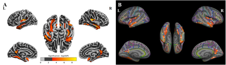
Subcortical ischemic vascular disease (SIVD) is a major cause of vascular cognitive impairment (CI) and features extensive atrophy in the cerebral cortex. We aimed to test the hypothesis that cognitive deficits in SIVD are linked to decreased cortical thickness in specific brain regions, which may constitute neuroimaging biomarkers of CI. Sixty-seven SIVD patients without (SIVD-NC, n = 35) and with (SIVD-CI, n = 32) CI and a group of healthy controls (HCs, n = 36) underwent structural magnetic resonance imaging (MRI) and cognitive functional assessments. FreeSurfer was used to preprocess structural MRI data and to calculate and compare cortical thickness. The correlation between cortical thickness and cognitive scores was examined in SIVD patients. Significantly altered cortical thickness in the bilateral insula, middle and inferior temporal lobes, precuneus, and medial temporal lobe (MTL) was identified among the three groups (p < 0.05, Monte Carlo simulation corrected). Post hoc results showed significantly decreased thickness in the bilateral insula and temporal lobe in SIVD-NC and SIVD-CI patients compared with HCs. However, the areas with reduced cortical thickness were larger in SIVD-CI than SIVD-NC patients. SIVD-CI patients had significantly reduced thickness in the bilateral precuneus and left MTL (Bonferroni corrected) compared with SIVD-NC patients when we extracted the mean thickness for each region of interest. In SIVD patients, the thicknesses of the left MTL and bilateral precuneus were positively correlated with immediate recall in the memory test. SIVD might lead to extensive cerebral cortical atrophy, while atrophy in the MTL and precuneus might be associated with memory deficits.
Keywords: cerebral small vessel disease; cognition; cortical thickness; magnetic resonance imaging; memory.
Copyright © 2021 Chen, Song, Cheng, Wang, Liu, He and Luo.
Conflict of interest statement
The authors declare that the research was conducted in the absence of any commercial or financial relationships that could be construed as a potential conflict of interest.
Figures


Similar articles
-
Study of gray matter atrophy pattern with subcortical ischemic vascular disease-vascular cognitive impairment no dementia based on structural magnetic resonance imaging.Front Aging Neurosci. 2023 Feb 6;15:1051177. doi: 10.3389/fnagi.2023.1051177. eCollection 2023. Front Aging Neurosci. 2023. PMID: 36815175 Free PMC article.
-
Unraveling the link: white matter damage, gray matter atrophy and memory impairment in patients with subcortical ischemic vascular disease.Front Neurosci. 2024 Feb 1;18:1355207. doi: 10.3389/fnins.2024.1355207. eCollection 2024. Front Neurosci. 2024. PMID: 38362024 Free PMC article.
-
Relationships Between Memory Impairments and Hippocampal Structure in Patients With Subcortical Ischemic Vascular Disease.Front Aging Neurosci. 2022 Apr 18;14:823535. doi: 10.3389/fnagi.2022.823535. eCollection 2022. Front Aging Neurosci. 2022. PMID: 35517055 Free PMC article.
-
Association of cortical macrostructural and microstructural changes with cognitive performance and gene expression in subcortical ischemic vascular disease patients with cognitive impairment.Brain Res Bull. 2025 Mar;222:111239. doi: 10.1016/j.brainresbull.2025.111239. Epub 2025 Feb 3. Brain Res Bull. 2025. PMID: 39909351
-
Altered Neurovascular Coupling in Subcortical Ischemic Vascular Disease.Front Aging Neurosci. 2021 May 12;13:598365. doi: 10.3389/fnagi.2021.598365. eCollection 2021. Front Aging Neurosci. 2021. PMID: 34054499 Free PMC article.
Cited by
-
Unraveling the Potential of EphA4: A Breakthrough Target and Beacon of Hope for Neurological Diseases.Cell Mol Neurobiol. 2023 Oct;43(7):3375-3391. doi: 10.1007/s10571-023-01390-0. Epub 2023 Jul 21. Cell Mol Neurobiol. 2023. PMID: 37477786 Free PMC article. Review.
-
Greater socioenvironmental risk factors and higher chronic pain stage are associated with thinner bilateral temporal lobes.Brain Behav. 2023 Dec;13(12):e3330. doi: 10.1002/brb3.3330. Epub 2023 Nov 20. Brain Behav. 2023. PMID: 37984835 Free PMC article.
-
Altered static and dynamic indices of intrinsic brain activity in patients with subcortical ischemic vascular disease: a resting-state functional magnetic resonance imaging analysis.Neuroradiology. 2023 May;65(5):923-931. doi: 10.1007/s00234-023-03135-8. Epub 2023 Mar 9. Neuroradiology. 2023. PMID: 36892613
-
Study of gray matter atrophy pattern with subcortical ischemic vascular disease-vascular cognitive impairment no dementia based on structural magnetic resonance imaging.Front Aging Neurosci. 2023 Feb 6;15:1051177. doi: 10.3389/fnagi.2023.1051177. eCollection 2023. Front Aging Neurosci. 2023. PMID: 36815175 Free PMC article.
-
Aberrant pattern of regional cerebral blood flow in mild cognitive impairment: A meta-analysis of arterial spin labeling magnetic resonance imaging.Front Aging Neurosci. 2022 Sep 1;14:961344. doi: 10.3389/fnagi.2022.961344. eCollection 2022. Front Aging Neurosci. 2022. PMID: 36118708 Free PMC article.
References
LinkOut - more resources
Full Text Sources
Other Literature Sources