

COVID-19-Associated Neurological Manifestations: An Emerging Electroencephalographic Literature
- PMID: 33679425
- PMCID: PMC7933549
- DOI: 10.3389/fphys.2020.622466
COVID-19-Associated Neurological Manifestations: An Emerging Electroencephalographic Literature
Abstract
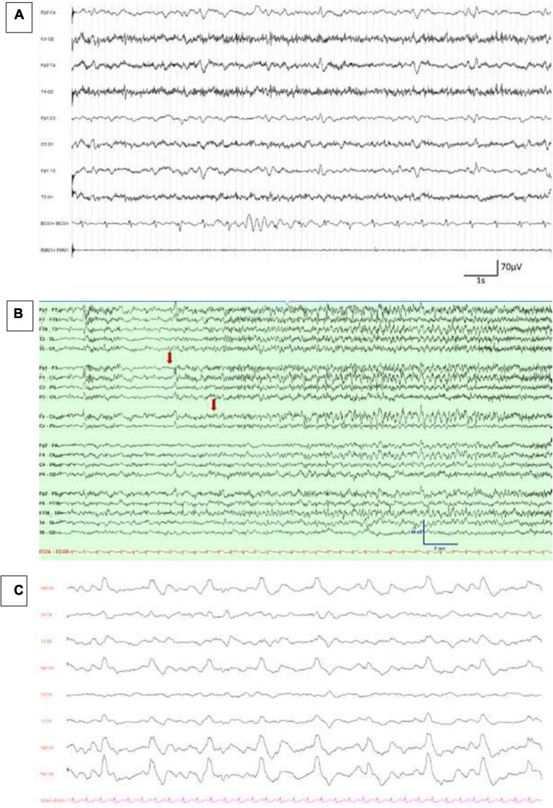
Severe acute respiratory syndrome coronavirus 2 (SARS-CoV-2) has spread worldwide since the end of year 2019 and is currently responsive for coronavirus infectious disease 2019 (COVID-19). The first reports considered COVID-19 as a respiratory tract disease responsible for pneumonia, but numerous studies rapidly emerged to warn the medical community of COVID-19-associated neurological manifestations, including encephalopathy at the acute phase and other postinfectious manifestations. Using standard visual analysis or spectral analysis, recent studies reported electroencephalographic (EEG) findings of COVID-19 patients with various neurological symptoms. Most EEG recordings were normal or revealed non-specific abnormalities, such as focal or generalized slowing, interictal epileptic figures, seizures, or status epilepticus. Interestingly, novel EEG abnormalities over frontal areas were also described at the acute phase. Underlying mechanisms leading to brain injury in COVID-19 are still unknown and matters of debate. These frontal EEG abnormalities could emphasize the hypothesis whereby SARS-CoV-2 enters the central nervous system (CNS) through olfactory structures and then spreads in CNS via frontal lobes. This hypothesis is reinforced by the presence of anosmia in a significant proportion of COVID-19 patients and by neuroimaging studies confirming orbitofrontal abnormalities. COVID-19 represents a new viral disease characterized by not only respiratory symptoms but also a systemic invasion associated with extra-respiratory signs. Neurological symptoms must be the focus of our attention, and functional brain evaluation with EEG is crucial, in combination with anatomical and functional brain imaging, to better understand its pathophysiology. Evolution of symptoms together with EEG patterns at the distance of the acute episode should also be scrutinized.
Keywords: COVID-19; EEG; SARS-CoV-2; coronavirus; encephalopathy; neurophysiology.
Copyright © 2021 Vellieux, Sonneville, Vledouts, Jaquet, Rouvel-Tallec and d’Ortho.
Conflict of interest statement
The authors declare that the research was conducted in the absence of any commercial or financial relationships that could be construed as a potential conflict of interest.
Figures
References
Publication types
LinkOut - more resources
Full Text Sources
Other Literature Sources
Miscellaneous