. 2021 Feb 18:12:635684.
doi: 10.3389/fmicb.2021.635684. eCollection 2021.

The Cysteine Protease MaOC1, a Prokaryotic Caspase Homolog, Cleaves the Antitoxin of a Type II Toxin-Antitoxin System

Affiliations

The Cysteine Protease MaOC1, a Prokaryotic Caspase Homolog, Cleaves the Antitoxin of a Type II Toxin-Antitoxin System

Marina Klemenčič et al. Front Microbiol. .

Abstract

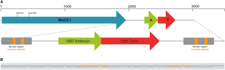
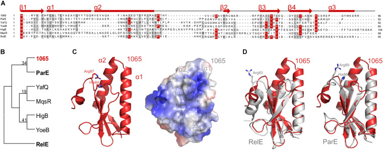
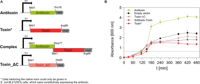
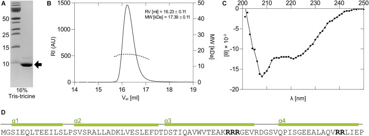
The bloom-forming cyanobacterium Microcystis aeruginosa is known for its global distribution and for the production of toxic compounds. In the genome of M. aeruginosa PCC 7806, we discovered that the gene coding for MaOC1, a caspase homolog protease, is followed by a toxin-antitoxin module, flanked on each side by a direct repeat. We therefore investigated their possible interaction at the protein level. Our results suggest that this module belongs to the ParE/ParD-like superfamily of type II toxin-antitoxin systems. In solution, the antitoxin is predominantly alpha-helical and dimeric. When coexpressed with its cognate toxin and isolated from Escherichia coli, it forms a complex, as revealed by light scattering and affinity purification. The active site of the toxin is restricted to the C-terminus of the molecule. Its truncation led to normal cell growth, while the wild-type form prevented bacterial growth in liquid medium. The orthocaspase MaOC1 was able to cleave the antitoxin so that it could no longer block the toxin activity. The most likely target of the protease was the C-terminus of the antitoxin with two sections of basic amino acid residues. E. coli cells in which MaOC1 was expressed simultaneously with the toxin-antitoxin pair were unable to grow. In contrast, no effect on cell growth was found when using a proteolytically inactive MaOC1 mutant. We thus present the first case of a cysteine protease that regulates the activity of a toxin-antitoxin module, since all currently known activating proteases are of the serine type.

Keywords: Microcystis aeruginosa; ParE/ParD; RelE/ParE; metacaspase; programmed cell death; protease; regulated cell death; toxin-antitoxin system.


Conflict of interest statement

The authors declare that the research was conducted in the absence of any commercial or financial relationships that could be construed as a potential conflict of interest.

Figures

FIGURE 1
FIGURE 1
Genomic context of the orthocaspase MaOC1 and the 1065–1067 toxin-antitoxin pair. Genes are shown in reverse orientation compared to the original sequence for clarity. (A) In Microcystis aeruginosa PCC 7806, the gene encoding the orthocaspase MaOC1 (in blue) is followed by a putative toxin-antitoxin (TA) gene pair (in red and green, respectively). The MaOC1 catalytic dyad, which is formed by His110 and Cys169, is denoted. Downstream and upstream of the TA pair, identical border repeats (in gray) are found, each containing a pair of inverted repeats (in orange). (B) The nucleotide sequence of the inverted repeat is shown as part of the border region.
FIGURE 2
FIGURE 2
Classification of the 1065 toxin. (A) Sequence alignment of the 1065 toxin with representatives of other RelE/ParE toxins (NCBI IDs: ParE, NP_310308; YafQ, NP_414760; YoeB, NP_707909; HigB, NP_311992; MqsR, NP_417494; RelE, NP_416081). Identical residues are colored red and similar amino acids are shaded gray with a threshold of 50% for coloration. Predicted secondary structures of the 1065 toxin are shown above the alignment. The sequence alignment was performed with PROMALS and the image was generated with BioEdit. (B) The unrooted phylogenetic tree of selected toxin representatives. The tree was constructed using the Neighbor-Joining method of the MEGA software (version 6) and is based on the alignment shown in panel A. The 1065 toxin is shown in red, while ParE and RelE toxins, whose structures are shown in panel D, are shown in bold. (C) Homology model of the 1065 toxin in cartoon representation (left) and its surface potential (right; where red shows the acidic surface potential and blue shows the basic surface potential). The models were predicted by I- TASSER software. (D) Overlay of the 1065 toxin model (shown in red) and the RelE (PDB: 4FXE) (left) or ParE (PDB: 5CW7) toxins (shown in gray) (right). Basic amino acid residues known to be responsible for toxic activity are shown as sticks. All images were generated with PyMOL (DeLano Scientific).
FIGURE 3
FIGURE 3
Expression of the 1065 (toxin) and 1067 (antitoxin) genes in E. coli BL21(DE3) cells. (A) We constructed four pET28b(+)-based vectors that, under the IPTG-inducible T7 promoter, directed the expression of the native antitoxin, the native toxin, the native TA pair (complex) or the C-terminally truncated toxin in E. coli BL21(DE3) cells. All genes were inserted in frame with the 3’ His-tag coding sequence. (B) The growth of E. coli BL21(DE3) cells at 37°C and shaken at 200 rpm was monitored for 480 min by measuring the optical density at 600 nm. The expression of genes under the T7 promoter was induced by 1 mM IPTG when OD600 reached about 0.6 (dotted line). Cells containing the pET28_toxin plasmid were co-transformed with the pSB1C3_antitoxin plasmid, allowing constitutive expression of the antitoxin under a strong promoter and a strong ribosome binding site. Cells transformed with the empty pET28b(+) vector were used as controls. Although no gene was under the regulation of the T7 promoter, IPTG was also added at the indicated time point.
FIGURE 4
FIGURE 4
Expression and properties of the 1067 antitoxin. (A) The 1067 antitoxin was overexpressed in E. coli as a C-terminally His-tagged protein and isolated by nickel-affinity chromatography. It migrated on a 16% Tris-tricine gel with an approximate molecular weight of slightly less than 10 kDa (black arrow). (B) Right and Low Light Scattering (RALS/LALS) showed that the antitoxin in solution was mostly present as a molecule with an approximate size of 17.4 kDa, indicating the formation of a dimeric structure. (C) The CD spectrum of the 1067 antitoxin in 20 mM HEPES, pH 7.4, showed that at 25°C it exhibited predominantly α-helical structure. This was further confirmed by secondary and tertiary structure prediction models (D), which predicted the folding of the protein into four α-helices. The clusters with two or more Arg residues are marked in bold.
FIGURE 5
FIGURE 5
Expression and multimeric form of the 1065–1067 toxin-antitoxin pair. (A) Right and Low Light Scattering (RALS/LALS) showed that in solution, the antitoxin-toxin complex was mostly present as a molecule with an approximate size of 27 kDa, indicating the formation of a heterotrimer, in which two molecules of the antitoxin bind to one molecule of the toxin. (B) 16% Tris-tricine polyacrylamide gel stained with the Coommasie Brilliant Blue dye. The toxin-antitoxin pair was expressed from the native locus arrangement, but with a His-tag at the C-terminus of the toxin and isolated by nickel affinity chromatography. The expected mass is 12.5 kDa for the His-tag labeled toxin and 8.4 kDa for the antitoxin.
FIGURE 6
FIGURE 6
Proteolytic cleavage of the antitoxin or the TA complex with the orthocaspase MaOC1. (A) The 1067 antitoxin was incubated in the presence of a wild-type MaOC1 or its proteolytically inactive variant MaOC1_C169A in the mass ratios given for 2 h at room temperature. The reaction mixtures were then electrophoresed on a 16% Tris-tricine gel and stained with Coommasie Brilliant Blue. The arrow represents the expected size of the full-length C-terminally tagged antitoxin (9.5 kDa). (B) Reactions containing the antitoxin with MaOC1 (ratio 1:50) were stopped at various times, loaded onto the 16% Tris-tricine gel and stained with Coommasie Brilliant Blue after electrophoresis. (C) The antitoxin (A) or the complex (AT) were incubated for 1 h in the absence or presence of MaOC1 orthocaspase (mass ratio 1:100 protein:protease) and applied in duplicates to the 16% Tris-tricine gel. After electrophoresis, the gel was stained with Coommasie Brilliant Blue. All cleavage assays were performed in 20 mM HEPES, pH 7.4, 150 mM NaCl and at room temperature.
FIGURE 7
FIGURE 7
Growth of BL21(DE3) E. coli cells expressing TA operon or/and MaOC1 under constitutive and inducible conditions. BL21DE3 E. coli cells were transformed either with a pSB1C3_empty (A) or a pSB1C3_toxin_antitoxin (B) plasmid. Those cells were further used for co-transformation with pET28_MaOC1_WT/C169A plasmid (T7_MaOC1_WT or T7_MaOC1_C169A). The cells containing the respective plasmids were cultured in liquid LB media supplemented with appropriate antibiotics (C, chloramphenicol; K, kanamycin; or both). Overnight cultures diluted to an initial OD600 of about 0.05 (except for const_ TA/T7_MaOC1_WT where overnight culture was used) were grown in the respective media at 37°C and OD600 was measured at the indicated times. For the expression of genes under the T7 promoter/lac operator, IPTG was added to the cultures at 1 mM final concentration (marked with the dotted line).

Similar articles

Cited by

References

    1. Aakre C. D., Herrou J., Phung T. N., Perchuk B. S., Crosson S., Laub M. T. (2015). Evolving new protein-protein interaction specificity through promiscuous intermediates. Cell 163 594–606. 10.1016/j.cell.2015.09.055 - DOI - PMC - PubMed
    1. Ames J. R., Muthuramalingam M., Murphy T., Najar F. Z., Bourne C. R. (2019). Expression of different ParE toxins results in conserved phenotypes with distinguishable classes of toxicity. Microbiol. Open 8:e902. 10.1002/mbo3.902 - DOI - PMC - PubMed
    1. Asplund-Samuelsson J., Bergman B., Larsson J. (2012). Prokaryotic caspase homologs: phylogenetic patterns and functional characteristics reveal considerable diversity. PLoS One 7:e49888. 10.1371/journal.pone.0049888 - DOI - PMC - PubMed
    1. Brown B. L., Lord D. M., Grigoriu S., Peti W., Page R. (2013). The Escherichia coli toxin MqsR destabilizes the transcriptional repression complex formed between the antitoxin MqsA and the mqsRA operon promoter. J. Biol. Chem. 288 1286–1294. 10.1074/jbc.M112.421008 - DOI - PMC - PubMed
    1. Brzozowska I., Zielenkiewicz U. (2013). Regulation of toxin-antitoxin systems by proteolysis. Plasmid 70 33–41. 10.1016/j.plasmid.2013.01.007 - DOI - PubMed

LinkOut - more resources