. 2021 Feb 17:12:632606.
doi: 10.3389/fimmu.2021.632606. eCollection 2021.

Inhibition of NLRP3 Inflammasome Activation and Pyroptosis in Macrophages by Taraxasterol Is Associated With Its Regulation on mTOR Signaling

Affiliations

Inhibition of NLRP3 Inflammasome Activation and Pyroptosis in Macrophages by Taraxasterol Is Associated With Its Regulation on mTOR Signaling

Fan Yang et al. Front Immunol. .

Abstract

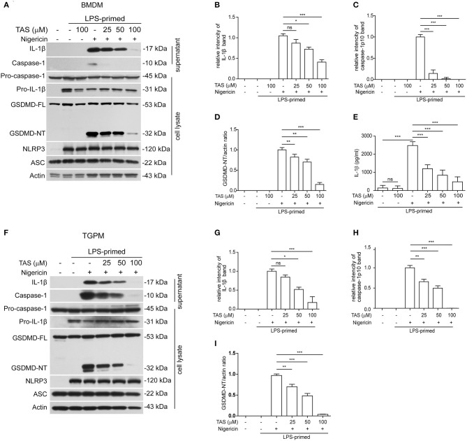
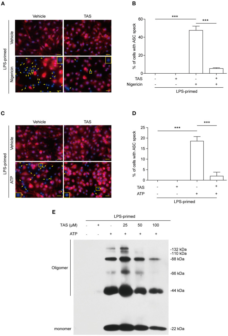
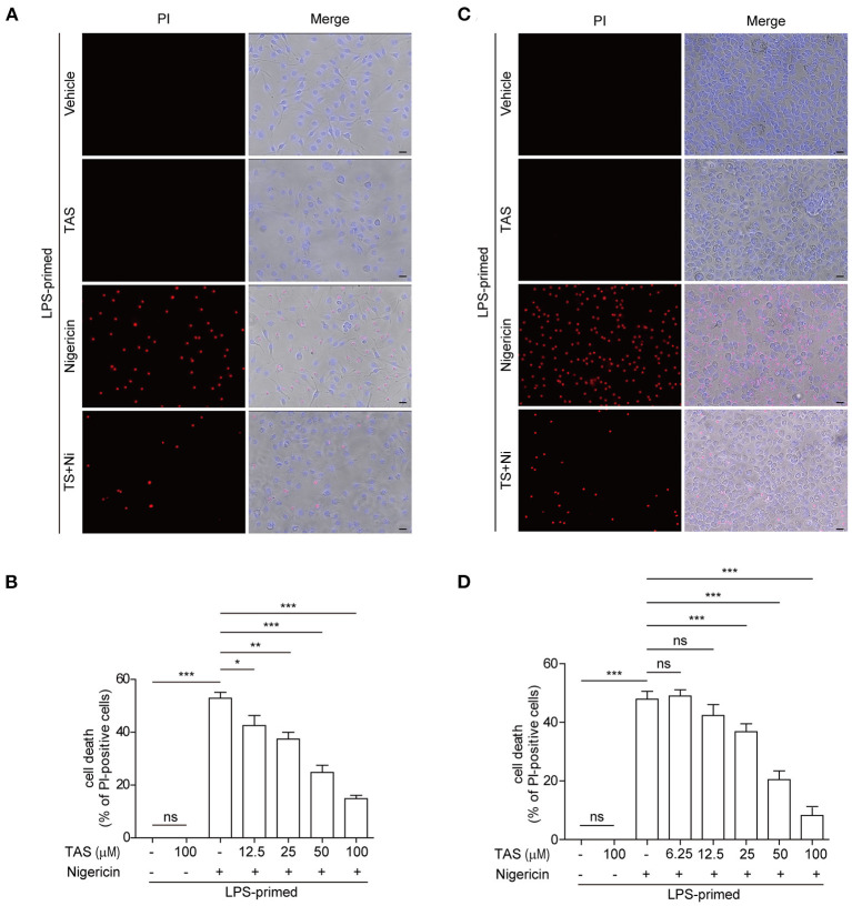
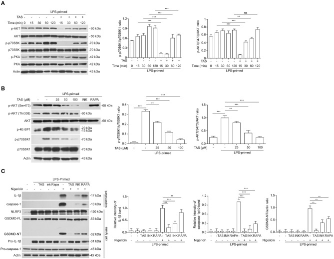
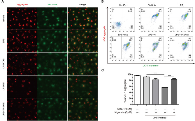
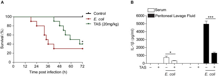
Taraxasterol (TAS) is an active ingredient of Dandelion (Taraxacum mongolicum Hand. -Mazz.), a medicinal plant that has long been used in China for treatment of inflammatory disorders. But the underlying mechanism for its therapeutic effects on inflammatory disorders is not completely clear. Inflammasome activation is a critical step of innate immune response to infection and aseptic inflammation. Among the various types of inflammasome sensors that has been reported, NLR family pyrin domain containing 3 (NLRP3) is implicated in various inflammatory diseases and therefore has been most extensively studied. In this study, we aimed to explore whether TAS could influence NLPR3 inflammasome activation in macrophages. The results showed that TAS dose-dependently suppressed the activation of caspase-1 in lipopolysaccharide (LPS)-primed murine primary macrophages upon nigericin treatment, resulting in reduced mature interleukin-1β (IL-1β) release and gasdermin D (GSDMD) cleavage. TAS greatly reduced ASC speck formation upon the stimulation of nigericin or extracellular ATP. Consistent with reduced cleavage of GSDMD, nigericin-induced pyroptosis was alleviated by TAS. Interestingly, TAS time-dependently suppressed the mechanistic target of rapamycin (mTOR) complex 1 (mTORC1) and mTORC2 signaling induced by LPS priming. Like TAS, both INK-128 (inhibiting both mTORC1 and mTORC2) and rapamycin (inhibiting mTORC1 only) also inhibited NLRP3 inflammasome activation, though their effects on mTOR signaling were different. Moreover, TAS treatment alleviated mitochondrial damage by nigericin and improved mouse survival from bacterial infection, accompanied by reduced IL-1β levels in vivo. Collectively, by inhibiting the NLRP3 inflammasome activation, TAS displayed anti-inflammatory effects likely through regulation of the mTOR signaling in macrophages, highlighting a potential action mechanism for the anti-inflammatory activity of Dandelion in treating inflammation-related disorders, which warrants further clinical investigation.

Keywords: ASC speck; NLRP3 inflammasome; dandelion; mTOR; taraxasterol.


Conflict of interest statement

The authors declare that the research was conducted in the absence of any commercial or financial relationships that could be construed as a potential conflict of interest.

Figures

Figure 1
Figure 1
Taraxasterol (TAS) dose-dependently suppressed nigericin-induced activation of NLRP3 inflammasome in murine macrophages. Bone marrow-derived macrophages (BMDMs) (A–E) or thioglycollate (TG)-elicited peritoneal macrophages (TGPMs) (F–I) were primed with LPS (0.5 μg/ml) for 4 h and then pretreated with indicated concentrations of taraxasterol (TAS) for 1 h, followed by stimulation with nigericin (3 μM for BMDMs and 2 μM for TGPMs) for 1 h. (A,F) Western blot analysis of the expression levels of indicated proteins in the culture supernatants and cell lysates. (B,C,G,H) Histograms show the relative intensity of capase-1p10 (10 kDa) or mature IL-1β (17 kDa) bands in supernatants, with the intensity of nigericin group being set to 1.0, respectively. (D,I) Relative GSDMD_NT levels in the cell lysates. (E) The concentration of soluble IL-1β in the culture supernatants were detected by cytometric bead array (CBA) assay. Data were analyzed using the non-parametric Mann–Whitney U-test, which are shown as mean ± SD (n = 3). *P < 0.05; **P < 0.01; ***P < 0.001; ns, not significant.
Figure 2
Figure 2
TAS inhibited ASC speck formation in macrophages. BMDMs were primed with LPS (0.5 μg/ml) for 4 h and then pretreated with TAS (100 μM) for 1 h, followed by stimulation with nigericin (3 μM) for 1 h (A,B) or ATP (2 mM) for 30 min (C,D). (A,C) Representative immunofluorescence images showing ASC (red) subcellular distribution. Nuclei were stained with Hoechst 33342 (blue). The images for ASC and nuclei were captured, respectively, and merged together. Yellow arrows indicate ASC specks and the insets show the cell with an ASC speck. (B,D) Histograms show the percentage of cells with an ASC speck relative to total cells from 5 randomly chosen fields. Data are shown as mean ± SD (n = 5). ***P < 0.001; ns, not significant; scale bars, 20 μm. ASC oligomerization after ATP stimulation was assayed by Western blotting (E).
Figure 3
Figure 3
TAS dose-dependently suppressed nigericin-induced pyroptosis in macrophages. BMDMs (A,B) were treated as in Figure 1A and TGPMs (C,D) were treated as in Figure 1E. (A,C) After TAS and nigericin (Ni) treatment, BMDMs and TGPMs were stained with 2 μg/ml propidium iodide (PI) (red; staining dead cells) and 5 μg/ml Hoechst 33342 (blue; staining all cells) for 10 min, and then observed by fluorescent microscopy. Bright-field images are also shown in merged ones (here images of TAS = 100 μM are shown). (B,D) The percentage of cell death is defined as PI-positive cells to all cells (Hoechst 33342-positive) in 5 random fields. Data are shown as mean ± SD (n = 5). *P < 0.05; **P < 0.01; ***P < 0.001; ns, not significant; scale bars, 20 μm.
Figure 4
Figure 4
TAS inhibited mTOR signaling in LPS-primed macrophages. LPS-primed BMDMs were primed with 0.5 μg/ml LPS for 4h, followed by treatment with 100 μM TAS for indicated time lengths (A), or indicated concentration of TAS for 15 min (B), or 100 μM TAS, 20 nM rapamycin (RAPA), 30 nM INK-128 (INK), respectively (C). The levels of indicated proteins in the cell lysates were analyzed by Western blotting. Actin was added as loading control. Data are shown as mean ± SD (n = 5). **P < 0.01; ***P < 0.001; ns, not significant.
Figure 5
Figure 5
Taraxasterol alleviates mitochondrial damage induced by nigericin. Mouse BMDMs were first primed with 0.5 μg/ml LPS for 4 h, then treated with 100 μM taraxasterol (TAS) for 1 h, and finally stimulated with 5 μM nigericin for 1 h. Mitochondrial membrane potential was assayed with a JC-1 kit according to the instruction of the manufacturer. JC-1 aggregates and monomers were observed by fluorescent microscopy (A) and evaluated by flow cytometry (B); the percentages of cells with JC-1 aggregates are shown (C). Data are shown as mean ± SD (n = 3). ***P < 0.001.
Figure 6
Figure 6
Taraxasterol increases mouse survival from bacterial infection. Female C57BL/6 mice were administered intragastrically (i.g.) with TAS or vehicle (2% Tween-80 in PBS); 3 h later, the mice were injected with freshly prepared viable E. coli (2 × 10⁹ CFU/mouse in 0.3 ml PBS) into the peritoneal cavity of each mouse; One hour after bacterial injection, the mice were given with a same dose of TAS again (i.g.). (A) Mouse survival was observed and recorded every 6 h for 3 consecutive days (n = 10). *P < 0.05, TAS (20 mg/kg) group vs. E. coli group. (B) In a parallel experiment, mouse serum and peritoneal lavage fluid was collected at 8 h post bacterial infection. IL-1β in the serum and peritoneal lavage fluid, respectively, were evaluated by CBA assay (n = 5).*P < 0.05; ***P < 0.001.

Similar articles

Cited by

References

    1. Chauhan D, Vande Walle L, Lamkanfi M. Therapeutic modulation of inflammasome pathways. Immunol Rev. (2020) 297:123–38. 10.1111/imr.12908 - DOI - PMC - PubMed
    1. Rai RC. Host inflammatory responses to intracellular invaders: review study. Life Sci. (2020) 240:117084. 10.1016/j.lfs.2019.117084 - DOI - PubMed
    1. Swanson KV, Deng M, Ting JP. The NLRP3 inflammasome: molecular activation and regulation to therapeutics. Nat Rev Immunol. (2019) 19:477–89. 10.1038/s41577-019-0165-0 - DOI - PMC - PubMed
    1. Guo H, Callaway JB, Ting JP. Inflammasomes: mechanism of action, role in disease, and therapeutics. Nat Med. (2015) 21:677–87. 10.1038/nm.3893 - DOI - PMC - PubMed
    1. Coll RC, Robertson AA, Chae JJ, Higgins SC, Munoz-Planillo R, Inserra MC, et al. . A small-molecule inhibitor of the NLRP3 inflammasome for the treatment of inflammatory diseases. Nat Med. (2015) 21:248–55. 10.1038/nm.3806 - DOI - PMC - PubMed

Publication types

MeSH terms

LinkOut - more resources