Review
. 2021 Feb 18:12:639902.
doi: 10.3389/fimmu.2021.639902. eCollection 2021.

Immunological Impact of Intestinal T Cells on Metabolic Diseases

Affiliations
Review

Immunological Impact of Intestinal T Cells on Metabolic Diseases

Haiyan Zhou et al. Front Immunol. .

Erratum in

Abstract

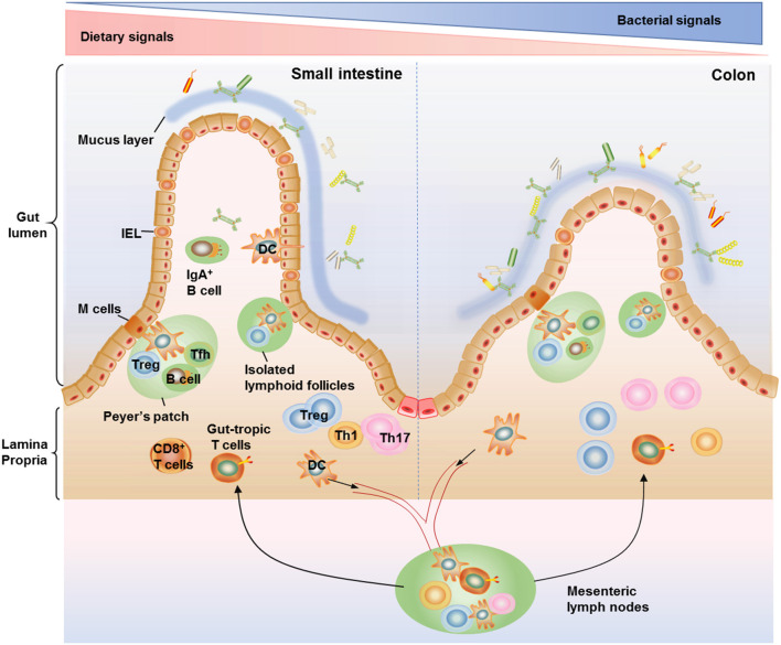
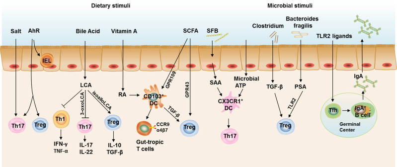
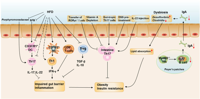
Emerging evidence accumulated over the past several years has uncovered intestinal CD4+ T cells as an essential mediator in modulating intestinal immunity in health and diseases. It has also been increasingly recognized that dietary and microbiota-derived factors play key roles in shaping the intestinal CD4+ T-cell compartment. This review aims to discuss the current understanding on how the intestinal T cell immune responses are disturbed by obesity and metabolic stress. In addition, we review how these changes influence systemic metabolic homeostasis and the T-cell-mediated crosstalk between gut and liver or brain in the progression of obesity and its related diseases. Lastly, we highlight the potential roles of some drugs that target intestinal T cells as a therapeutic treatment for metabolic diseases. A better understanding of the interaction among metabolites, bacterial signals, and T cell immune responses in the gut and their roles in systemic inflammation in metabolic tissues should shed new light on the development of effective treatment of obesity and related disorders.

Keywords: T cells; dietary signals; intestine; microbiota; obesity.


Conflict of interest statement

The authors declare that the research was conducted in the absence of any commercial or financial relationships that could be construed as a potential conflict of interest.

Figures

Figure 1
Figure 1
Anatomical distribution of intestinal T cells. T cells distributed within the small and large intestine are frequently arranged within the gut-associated lymphoid tissue (GALT), which are composed of organized lymphoid tissues including mesenteric lymph nodes, Peyer's patches, and isolated lymphoid follicles (ILFs) as well as more diffusely scattered lymphocytes including the lamina propria lymphocytes and intraepithelial lymphocytes (IELs). IELs reside within the epithelium layer and are proximity to antigens in the gut lumen, making them components of the front line of immune defense against invading pathogens. There are more IELs in the small intestine compared with the colon. The majority of T cells in lamina propria are CD4+ T cells, with only a small population of CD8+ T cells. Among the CD4+ T cell subsets, Th17 cells and Treg cells are the most abundant cells in the gut, conferring protection against fungi and maintenance of intestinal homeostasis. The organization of Peyer's patches is comparable to that of lymph nodes, with large B cell follicles and T cell areas. Peyer's patches are in close contact with microfold cells (M cells) located in gut epithelium which can capture and transport antigens from the lumen to antigen-presenting cells such as dendritic cells (DCs) in the underlying Peyer's patches. DCs can also form transepithelial dendrites to directly sample luminal antigens and then emigrate to the mesenteric lymph nodes, where they activate naive T cells to induce gut-tropic T cells. The ILFs, with features similar to Peyer's patches, are distributed along the whole intestinal tract and believed to provide a complementary system for Peyer's patches.
Figure 2
Figure 2
Intestinal T cell immune responses under steady state. Th17 cells and Treg cells are the most abundant CD4+ T cells in the gut to maintain intestinal homeostasis. Excessive salt intake and AhR ligation both contribute to the increased Th17 cell polarization in the gut, while AhR ligation also required for the induction of Treg cells and maintenance of IELs. Bile acids are cholesterol-derived natural surfactants abundant in the mammalian gut. Lithocholic acid, a secondary metabolite of bile acids, impedes Th1 activation and two distinct derivatives of lithocholic acid, 3-oxo-lithocholic acid and isoallo-lithocholic acid, have been found to impair the differentiation of Th17 cells and increase the differentiation of Treg cells, respectively. CD103+ DCs represent the dominant DC population in the murine small intestinal lamina propria (SILP). CD103+ DCs induce the gut-homing receptors CC chemokine receptor (CCR)9 and α4β7 on responding T cells and Treg cell differentiation, both of which are dependent on signaling events initiated by the vitamin A metabolite, retinoic acid (RA). Short-chain fatty acids (SCFAs) could also induce Treg polarization directly through a receptor GPR43 on T cells or indirectly through a receptor GPR109 on CD103+ DCs. Segmented filamentous bacteria (SFB) induce serum amyloid A (SAA) production from gut epithelial cells and stimulates CX3CR1+ DCs to promote Th17 cell development in the gut, the latter process could also be induced by commensal organism-derived ATP stimulation. Clostridium strains provide an environment rich in TGF-β that induces IL-10-producing colonic Treg Cells. Bacteroides fragilis (B. fragilis) promotes the differentiation and function Treg via its immunomodulatory molecule polysaccharide A (PSA), where Toll-like receptor 2 (TLR2) signaling is required. TLR2 ligand could also stimulate Tfh development in the germinal center to facilitate IgA production by B cells. Taken together, IL-17 and IL-22 produced by Th17, IL-10 and TGF-β produced by Treg cells, and IgA in the gut lumen constitute an immune barrier in maintaining gut homeostasis in steady state.
Figure 3
Figure 3
The roles of intestinal T cell immune responses in the development of obesity. HFD feeding reduces the proportions and numbers of RORγt+Th17 and Treg cells and increases the proportion of IFN-γ+ Th1 cells and CD8+ T cells in the SILP. HFD feeding also enhances the ability of CD103+ DCs to induce Th1 differentiation and inhibits the ability of CX3CR1+ cells to induce Th17 differentiation. The reduction of Porphyromonadaceae and SFB in the ileum at the onset of HFD feeding could be responsible for the reduction of intestinal Th17 cells. The increased IFN-γ production by Th1 and CD8+T cells impairs the gut barrier function and induces intestinal inflammation, which is aggravated by decreased production of IL-17, IL-22, TGF-β, and IL-10 from Th17 and Treg cells. Depletion of vitamin A in obese mice further reduced the proportion of Th17 cells in the small intestine, leading to increased body weight gain and insulin resistance and the metabolic disorders could be ameliorated by adoptive transfer of in vitro-differentiated gut-tropic Th17 cells to obese mice. Increasing Th17 cell proportion or function in the intestine by pretreating mice with dextran sodium sulfate or IL-22 administration also prevents HFD-induced glucose intolerance and insulinemia. However, Rag1−/− mice adoptive transferred with T cells from RORγt−/− mice leads to increased body weight gain and insulin resistance when compared with those adoptive transferred with wild-type T cells. IgA is also important in maintaining metabolic homeostasis by modulating gut microbiota homeostasis, while T cell-specific MyD88 knockout results in abnormal IgA antibody responses and an altered microbial gut community, leading to more severe HFD- and age-associated metabolic disorders.

References

    1. Tartof S, Qian L, Hong V, Wei R, Nadjafi R, Fischer H, et al. . Obesity and mortality among patients diagnosed with COVID-19: results from an integrated Health Care Organization. Ann Int Med. (2020) 173:773–81. 10.7326/M20-3742 - DOI - PMC - PubMed
    1. Popkin BM, Du S, Green WD, Beck MA, Algaith T, Herbst CH, et al. . Individuals with obesity and COVID-19: a global perspective on the epidemiology and biological relationships. Obes Rev. (2020) 21:e13128. 10.1111/obr.13128 - DOI - PMC - PubMed
    1. Zhou H, Liu F. Regulation, communication, and functional roles of adipose tissue-resident CD4+ T cells in the control of metabolic homeostasis. Front Immunol. (2018) 9:1961. 10.3389/fimmu.2018.01961 - DOI - PMC - PubMed
    1. Field CS, Baixauli F, Kyle RL, Puleston DJ, Cameron AM, Sanin DE, et al. . Mitochondrial integrity regulated by lipid metabolism is a cell-intrinsic checkpoint for treg suppressive function. Cell Metab. (2020) 31:422–37.e5. 10.1016/j.cmet.2019.11.021 - DOI - PMC - PubMed
    1. Garidou L, Pomie C, Klopp P, Waget A, Charpentier J, Aloulou M, et al. . The gut microbiota regulates intestinal CD4 T cells expressing RORgammat and controls metabolic disease. Cell Metab. (2015) 22:100–12. 10.1016/j.cmet.2015.06.001 - DOI - PubMed

Publication types