

Multivalvular destruction as the primary presentation of aggressive infective endocarditis with subaortic valve membrane
- PMID: 33679959
- PMCID: PMC7931308
- DOI: 10.12669/pjms.37.2.2798
Multivalvular destruction as the primary presentation of aggressive infective endocarditis with subaortic valve membrane
Abstract
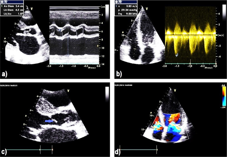
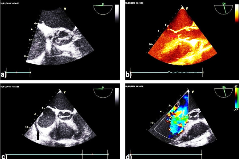
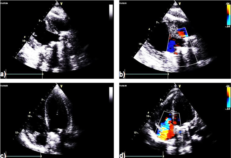
Multivalvular destruction may be a clinical manifestation of infective endocarditis (IE), which is a devastating infection of the heart either alone or superimposed with congenital subaortic membrane as in this case report. Here, we report a case of multivavular destruction with severe vegetation presented as a manifestation of infective endocarditis (IE) in a neglected case of 18-year-old male with previous rheumatic heart disease. Transesophageal echocardiography is an important imaging modality for diagnosis of superimposed aortic and heart lesions. Early necessary investigation and correct diagnosis is mandatory to prevent bad complications.
Keywords: Aortic regurgitation; Infective endocarditis; Mitral regurgitation; TEE; TTE.
Copyright: © Pakistan Journal of Medical Sciences.
Conflict of interest statement
Conflicts of interests: The authors have disclosed no conflicts of interest. The authors approved the publication, and take responsibility for its accuracy and integrity.
Figures
Similar articles
-
Transesophageal echocardiographic recognition of subaortic complications in aortic valve endocarditis. Clinical and surgical implications.Circulation. 1992 Aug;86(2):353-62. doi: 10.1161/01.cir.86.2.353. Circulation. 1992. PMID: 1638704
-
Transthoracic echocardiography (TTE): sufficiently sensitive screening test for native valve infective endocarditis (IE).Heart Lung. 2011 Jul-Aug;40(4):358-60. doi: 10.1016/j.hrtlng.2010.07.007. Epub 2011 Apr 11. Heart Lung. 2011. PMID: 21481468
-
Ventricular Septal Defect from Aortic Regurgitation Jet Lesion in Aortic Valve Infective Endocarditis.J Heart Valve Dis. 2016 Mar;25(2):150-152. J Heart Valve Dis. 2016. PMID: 27989057
-
Has transesophageal echocardiography changed the approach to patients with suspected or known infective endocarditis?Echocardiography. 1995 Nov;12(6):637-50. doi: 10.1111/j.1540-8175.1995.tb00857.x. Echocardiography. 1995. PMID: 10158101 Review.
-
Value and limitations of transesophageal echocardiography in infective endocarditis.Herz. 1993 Dec;18(6):341-60. Herz. 1993. PMID: 8307551 Review.
Cited by
-
Subaortic membrane in adult patients: retrospective study of clinical characteristics, outcomes, and echocardiographic associations and review of literature.Int J Cardiovasc Imaging. 2025 Aug 7. doi: 10.1007/s10554-025-03489-5. Online ahead of print. Int J Cardiovasc Imaging. 2025. PMID: 40773030
References
-
- Cahill TJ, Prendergast BD. Infective endocarditis. Lancet. 2016;387(10021):882–893. doi:10.1016/S0140-6736(15)00067-7. - PubMed
-
- Cahill TJ, Baddour LM, Habib G, Hoen B, Salaun E, Pettersson GB, et al. Challenges in Infective Endocarditis. J Am Coll Cardiol. 2017;69(3):325–344. doi:10.1016/j.jacc.2016.10.066. - PubMed
-
- Vincent LL, Otto CM. Infective Endocarditis:Update on Epidemiology Outcomes, and Management. Curr Cardiol Rep. 2018;20(10):86. doi:10.1007/s11886-018-1043-2. - PubMed
-
- Büchi A, Hoffmann M, Zbinden S, Atkinson A, Sendi P. The Duke minor criterion “predisposing heart condition”in native valve infective endocarditis - a systematic review. Swiss Med Wkly. 2018;148:w14675. doi:10.4414/smw.2018.14675. - PubMed
-
- Afonso L, Kottam A, Reddy V, Penumetcha A. Echocardiography in Infective Endocarditis:State of the Art. Curr Cardiol Rep. 2017;19(12):127. doi:10.1007/s11886-017-0928-9. - PubMed
Publication types
LinkOut - more resources
Full Text Sources
Other Literature Sources
Research Materials