Case Reports
. 2021 Feb 24;16(5):1037-1041.
doi: 10.1016/j.radcr.2021.02.012. eCollection 2021 May.

Ischial osteoid osteoma: A cause of persistent hip pain in an adolescent patient with bilateral femoroacetabular impingement

Affiliations
Case Reports

Ischial osteoid osteoma: A cause of persistent hip pain in an adolescent patient with bilateral femoroacetabular impingement

Alexander J Mortensen et al. Radiol Case Rep. .

Erratum in

Abstract

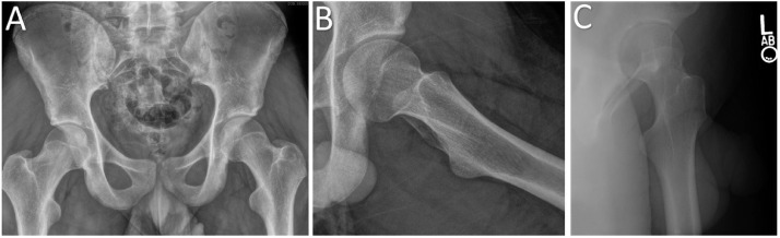
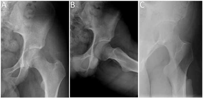
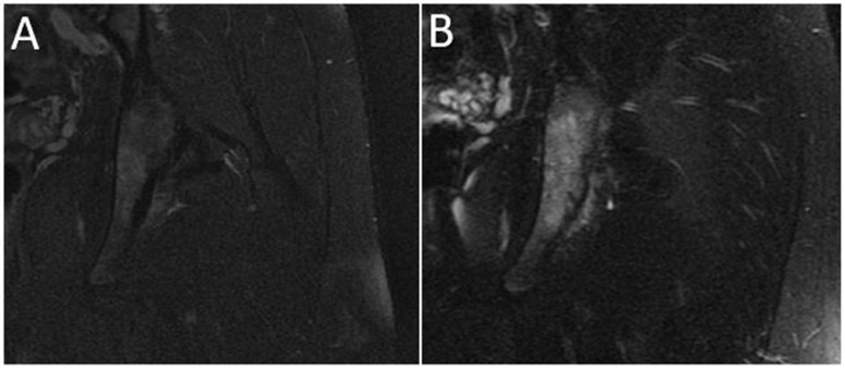
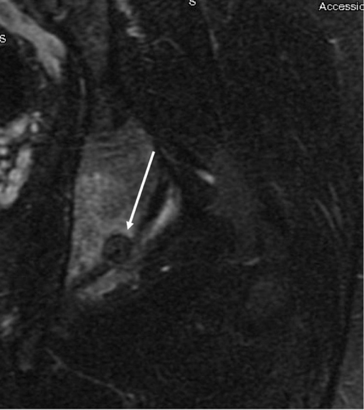
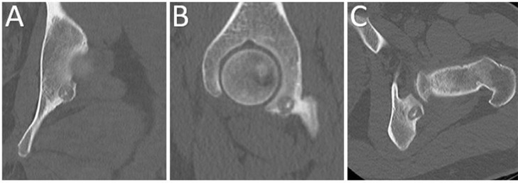
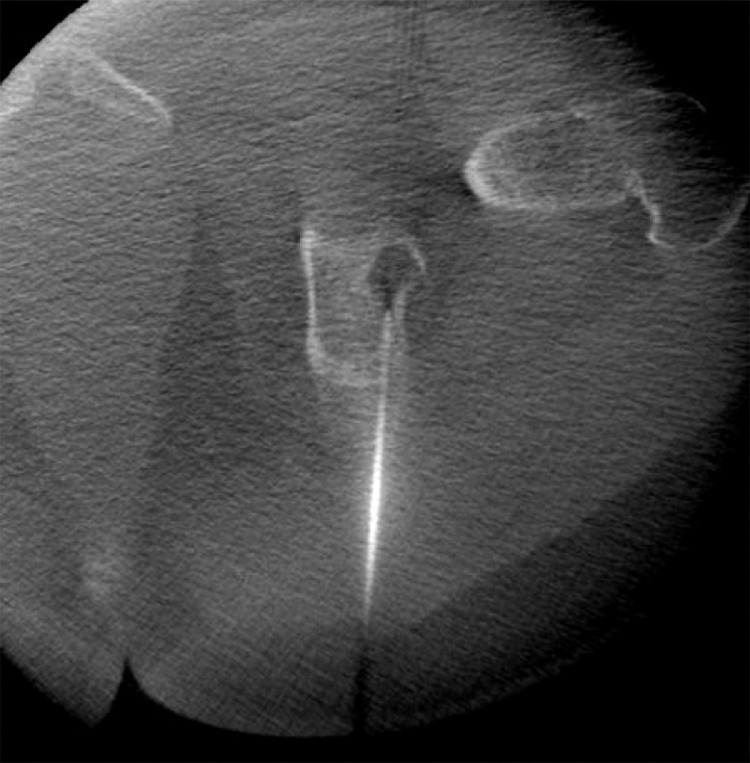
A 15-year-old boy presented with left-sided hip pain and imaging consistent with the diagnosis of femoroacetabular impingement. Following hip arthroscopy, which included an osteochondroplasty, labral repair, and capsular repair, the patient's anterior hip pain improved. However, his deep aching hip pain persisted until an ischial osteoid osteoma was identified and treated with radiofrequency ablation. At 3 years follow-up, the patient reports high satisfaction and minimal pain. We present this case to illustrate the importance of considering all potential causes of persistent hip pain following hip arthroscopy, including benign bone tumors which may be difficult to visualize on plain radiographs.

Keywords: Femoroacetabular impingement; Osteoid osteoma; Pediatric.


Figures

Fig. 1 –
Fig. 1
Radiographic evaluation prior to hip arthroscopy. Preoperative radiographic evaluation of the left hip in the setting of history and physical examination consistent with femoroacetabular impingement, consistent with CAM-type femoroacetabular impingement. (A) Anteroposterior (AP) pelvis radiograph, (B) Frog-leg lateral radiograph, (C) False profile radiograph.
Fig. 2 –
Fig. 2
Radiographic evaluation 7 months following femoral osteochondroplasty and labral repair. Postoperative radiographic evaluation of the left hip demonstrating adequate femoral osteochondroplasty. (A) Anteroposterior (AP) pelvis radiograph, (B) Frog-leg lateral radiograph, (C) false profile radiograph.
Fig. 3 –
Fig. 3
Comparison of the posterior-inferior acetabulum on magnetic resonance arthrography prior to and after surgery. Coronal T2-weighted fat saturated magnetic resonance images of the left hip showing the posterior-inferior acetabulum. (A) Magnetic resonance arthrogram obtained one month prior to left hip arthroscopy demonstrating normal findings. (B) Magnetic resonance arthrogram obtained 9 months following left hip arthroscopy demonstrating edema in the posterior-inferior acetabulum extending into the ischial tuberosity.
Fig. 4
Fig. 4
Bony lesion visualized on magnetic resonance arthrogram nine months following left hip arthroscopy. Coronal T2-weighted fat saturated magnetic resonance image of the left ischium, demonstrating an area of nidus formation with surrounding peritumoral edema (arrow).
Fig. 5
Fig. 5
Computed tomography of the left hip demonstrating a bony lesion of the ischium. Computed tomography scan of the left hip 10 months following left-sided hip arthroscopy. The osteoid osteoma is apparent in each image in the posterior-inferior acetabulum as a well-defined, lucent lesion with a nidus of central calcification. (A) Coronal slice through the ischium, (B) Sagittal slice through the medial femoral head and acetabulum, (C) Axial slice through the femoral neck and posterior-inferior acetabulum.
Fig. 6 –
Fig. 6
Radiofrequency ablation of the osteoid osteoma. Radiofrequency ablation probe positioned in the osteoid osteoma lesion of the left ischial tuberosity.

References

    1. Sardana V, Philippon MJ, De Sa D, Bedi A, Ye L, Simunovic N. Revision hip arthroscopy indications and outcomes: a systematic review. Arthroscopy. 2015;31(10):2047–2055. doi: 10.1016/j.arthro.2015.03.039. - DOI - PubMed
    1. Wylie JD, Beckmann JT, Maak TG, Aoki SK. Arthroscopic capsular repair for symptomatic hip instability after previous hip arthroscopic surgery. Am J Sports Med. 2016;44(1):39–45. doi: 10.1177/0363546515608162. - DOI - PubMed
    1. Spiker AM, Rotter B-Z, Chang B, Mintz DN, Kelly BT. Clinical presentation of intra-articular osteoid osteoma of the hip and preliminary outcomes after arthroscopic resection: a case series. J Hip Preserv Surg. 2018;5(1):88–99. doi: 10.1093/jhps/hnx042. - DOI - PMC - PubMed
    1. Banga K, Racano A, Ayeni OR, Deheshi B. Atypical hip pain: coexistence of femoroacetabular impingement (FAI) and osteoid osteoma. Knee Surg Sports Traumatol Arthrosc. 2015;23(5):1571–1574. doi: 10.1007/s00167-014-2985-6. - DOI - PubMed
    1. Atesok KI, Alman BA, Schemitsch EH, Peyser A, Mankin H. Osteoid osteoma and osteoblastoma. J Am Acad Orthop Surg. 2011;19(11):678–689. doi: 10.5435/00124635-201111000-00004. - DOI - PubMed

Publication types