

Endoscopic treatment of ectopic teeth in the maxillary sinus
- PMID: 33680324
- PMCID: PMC7920564
- DOI: 10.4317/jced.57905
Endoscopic treatment of ectopic teeth in the maxillary sinus
Abstract
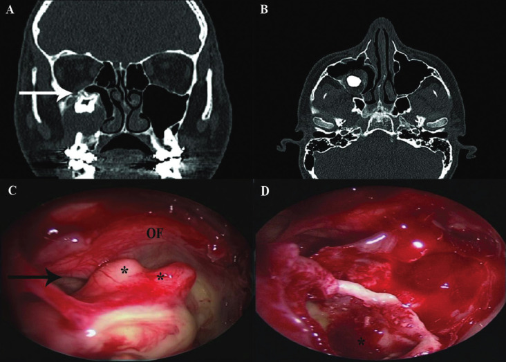
Background: Ectopic teeth in maxillary sinus is rare and are usually removed via sub-labial trans-canine fossa approach (SLCFA). The aim of our study was to present our experience with extraction of ectopic teeth in the maxillary sinus via transnasal endoscopic approach (TEA).
Material and methods: Rhinologists were asked to share their experience in the management of ectopic teeth in the maxillary sinus. Data were reviewed retrospectively.
Results: Eleven cases were reported in 10 patients from 2010 to 2019, six males and four females with a mean age of 33.5 +/-17 years (range 16 to 61). Seven patients complained of sinonasal symptoms, two were diagnosed incidentally during routine dental work-up, and one had oro-antral fistula. In eight patients, a cyst coexisted within the maxillary sinus. Teeth were located arbitrarily within the sinus. All cases were operated by TEA. One patient had self-limited periorbital emphysema, one had transient cheek numbness, and one had early post-operative bleeding that stopped after endoscopic cauterization. Long-term follow-up revealed good clinical outcomes.
Conclusions: Transnasal endoscopic removal of ectopic teeth from the maxillary sinus is a feasible and rational approach when SLCFA is contraindicated. Key words:Ectopic teeth, dentigerous cyst, endoscopic sinus surgery, Caldwell-Luc.
Copyright: © 2021 Medicina Oral S.L.
Conflict of interest statement
Conflicts of interest All authors declare no conflict of interest.
Figures

Similar articles
-
Transnasal endoscopic removal of an ectopic maxillary sinus tooth associated with a dentigerous cyst: A case report and a brief review.Int J Surg Case Rep. 2025 Mar;128:111049. doi: 10.1016/j.ijscr.2025.111049. Epub 2025 Feb 13. Int J Surg Case Rep. 2025. PMID: 39970609 Free PMC article.
-
Ectopic Tooth in the Roof of the Left Maxillary Sinus.Cureus. 2023 Dec 1;15(12):e49765. doi: 10.7759/cureus.49765. eCollection 2023 Dec. Cureus. 2023. PMID: 38164311 Free PMC article.
-
Endonasal endoscopic management of different cases of dentigerous cysts and ectopic teeth.J Surg Case Rep. 2021 Apr 19;2021(4):rjab099. doi: 10.1093/jscr/rjab099. eCollection 2021 Apr. J Surg Case Rep. 2021. PMID: 33897999 Free PMC article.
-
Endoscopic removal of ectopic sinonasal teeth: a systematic review.J Otolaryngol Head Neck Surg. 2019 Jul 5;48(1):30. doi: 10.1186/s40463-019-0353-8. J Otolaryngol Head Neck Surg. 2019. PMID: 31277707 Free PMC article.
-
Ectopic teeth in the maxillary sinus: A case report and literature review.Indian J Dent Res. 2018 Sep-Oct;29(5):667-671. doi: 10.4103/ijdr.IJDR_347_17. Indian J Dent Res. 2018. PMID: 30409951 Review.
Cited by
-
A Unique Case of Supernumerary Teeth Erupting Inside a Maxillary Sinus Osteoma.J Clin Med. 2024 Jul 11;13(14):4067. doi: 10.3390/jcm13144067. J Clin Med. 2024. PMID: 39064108 Free PMC article.
-
A Rare Case Report of a Large Dentigerous Cyst in the Maxillary Sinus Associated With an Ectopic Maxillary Third Molar.Case Rep Dent. 2025 Jul 3;2025:2436615. doi: 10.1155/crid/2436615. eCollection 2025. Case Rep Dent. 2025. PMID: 40642023 Free PMC article.
-
Endoscopically assisted removal of ectopic teeth in the floor of the orbit. Case report and latest literature review.J Clin Exp Dent. 2024 May 1;16(5):e639-e642. doi: 10.4317/jced.61371. eCollection 2024 May. J Clin Exp Dent. 2024. PMID: 38988746 Free PMC article.
-
Transinusal Pathway Removal of an Impacted Third Molar with an Unusual Approach: A Case Report and a Systematic Review of the Literature.Antibiotics (Basel). 2022 May 13;11(5):658. doi: 10.3390/antibiotics11050658. Antibiotics (Basel). 2022. PMID: 35625302 Free PMC article. Review.
-
Ectopic Upper Third Molar in Maxillary Sinus: A Case Report and Literature Review.Indian J Otolaryngol Head Neck Surg. 2022 Dec;74(Suppl 3):4718-4721. doi: 10.1007/s12070-021-03039-0. Epub 2022 Jan 18. Indian J Otolaryngol Head Neck Surg. 2022. PMID: 36742931 Free PMC article.
References
-
- Thesleff I, Nieminen P. Tooth morphogenesis and cell differentiation. Curr Opin Cell Biol. 1996;8:844–50. - PubMed
-
- Baykul T, Dogru H, Yasan H, Cina Aksoy M. Clinical impact of ectopic teeth in the maxillary sinus. Auris Nasus Larynx. 2006;33:277–81. - PubMed
-
- Buyukkurt MC, Omezli MM, Miloglu O. Dentigerous cyst associated with an ectopic tooth in the maxillary sinus: a report of 3 cases and review of the literature. Oral Surg Oral Med Oral Pathol Oral Radiol Endod. 2010;109:67–71. - PubMed
-
- Erkmen N, Olmez S, Onerci M. Supernumerary tooth in the maxillary sinus: case report. Aust Dent J. 1998;43:385–6. - PubMed
-
- Hasbini AS, Hadi U, Ghafari J. Endoscopic removal of an ectopic third molar obstructing the osteomeatal complex. Ear Nose Throat J. 2001;80:667–70. - PubMed
LinkOut - more resources
Full Text Sources
Other Literature Sources