

Delayed phases of contrast MRI, can it be valuable in multiple sclerosis active phase diagnosis?
- PMID: 33680386
- PMCID: PMC7911766
- DOI: 10.22088/cjim.11.4.432
Delayed phases of contrast MRI, can it be valuable in multiple sclerosis active phase diagnosis?
Abstract
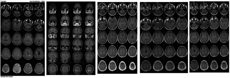
Background: Observing the enhancing plaques in magnetic resonance imaging (MRI) is one of the most valuable diagnostic modalities in confirming the diagnosis of multiple sclerosis (MS), its recurrence and for better detection of active disease. Since active lesions discovery can improve designating diffusion in time diagnosis of MS and controlling disease activity, and there is not any definite time for delay image acquisition, therefore, the aim of the current study was to evaluate the enhancement of MS plaques in different delayed phases.
Methods: In this interventional study, after receiving written consent, 40 MS patients with at least one enhancing plaque in a previous MRI were evaluated in Babol Ayatollah Rouhani Hospital. Gadolinium was injected to all patients at the dose of 0.1 mg/kg, and MRI was taken at 5 and 15 minutes. The results were analyzed using SPSS 23. A p<0.05 was considered as significant level.
Results: The mean of plaque signal intensity was 1190.20 and 1349.60 at 5 and 15 min, respectively, and this difference was significant (p<0.001). Moreover, the mean of plaque total size was 5.16 cm and 7.04 cm at 5 and 15 min with significant difference, respectively (p<0.001). The mean of plaque number was 1.92 and 2.58 at 5 and 15 min, respectively, which was significantly different (P<0.001).
Conclusion: The results indicated improvement in detection of MS plaques in images taken in the delayed phase compared to those in the early phase. The plaque intensity, size and number were significantly higher in the delayed phase (15 min), than early phase (5 min).
Keywords: Contrast M; Multiple sclerosis; Plaque count; Plaque enhancement; Plaque size.
Copyright © 2020, Babol University of Medical Sciences.
Figures
Similar articles
-
Evaluation of plaque detection and optimum time of enhancement in acute attack multiple sclerosis after contrast injection.Acta Radiol. 2014 Mar;55(2):218-24. doi: 10.1177/0284185113495831. Epub 2013 Aug 24. Acta Radiol. 2014. PMID: 23975149
-
Correlation between contrast enhanced plaques and plaque diffusion restriction and their signal intensities in FLAIR images in patients who admitted with acute symptoms of multiple sclerosis.J Med Imaging Radiat Sci. 2021 Mar;52(1):121-126. doi: 10.1016/j.jmir.2020.12.001. Epub 2021 Jan 11. J Med Imaging Radiat Sci. 2021. PMID: 33446443
-
Mitoxantrone: a review of its use in multiple sclerosis.CNS Drugs. 2004;18(6):379-96. doi: 10.2165/00023210-200418060-00010. CNS Drugs. 2004. PMID: 15089110 Review.
-
The relationship between enhanced plaques with Gadovist and Magnevist contrast brain magnetic resonance imaging and the neurological deficit in the acute phase of relapsing remitting multiple sclerosis.Iran J Neurol. 2012;11(2):42-6. Iran J Neurol. 2012. PMID: 24250860 Free PMC article.
-
[Magnetic resonance imaging in multiple sclerosis].Pathol Biol (Paris). 2000 Mar;48(2):151-61. Pathol Biol (Paris). 2000. PMID: 10815291 Review. French.
Cited by
-
Diffusion-Weighted Images and Contrast-Enhanced MRI in the Diagnosis of Different Stages of Multiple Sclerosis of the Central Nervous System.Cureus. 2023 Jul 10;15(7):e41650. doi: 10.7759/cureus.41650. eCollection 2023 Jul. Cureus. 2023. PMID: 37575819 Free PMC article.
References
-
- Ramagopalan SV, Sadovnick AD. Epidemiology of multiple sclerosis. Neurol Clin. 2011;29:207–17. - PubMed
-
- Wingerchuk DM, Lucchinetti CF, Noseworthy JH. Multiple sclerosis: current pathophysiological concepts. Lab Invest. 2001;81:263–81. - PubMed
-
- Orton SM, Herrera BM, Yee IM, et al. Sex ratio of multiple sclerosis in Canada: a longitudinal study. Lancet Neurol. 2006;5:932–6. - PubMed
-
- Filippi M, Rocca MA. MR imaging of multiple sclerosis. Radiology. 2011;259:659–81. - PubMed
LinkOut - more resources
Full Text Sources